
Page 85 - Read Online
P. 85
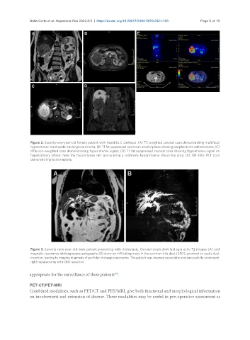
Della Corte et al. Hepatoma Res 2022;8:5  https://dx.doi.org/10.20517/2394-5079.2021.103  Page 5 of 15


                Figure 2. Seventy-one-year-old female patient with hepatitis C cirrhosis. (A) T2 weighted coronal scan demonstrating multifocal
                hyperintense intrahepatic cholangiocarcinoma; (B) T1 fat suppressed axial scan arterial phase showing peripheral rim enhancement; (C)
                diffusion-weighted scan demonstrating hyperintense signal; (D) T1 fat suppressed coronal scan showing hypointense signal on
                hepatobiliary phase: note the hypointense rim surrounding a relatively hyperintense cloud-like area; (E) 18F-FDG PET scan
                demonstrating lesions uptake.


                Figure 3. Seventy-nine-year-old male patient presenting with cholestasis. Coronal single shot fast spin echo T2 images (A) and
                magnetic resonance cholangiopancreatography (B) show an infiltrating mass in the common bile duct (CBD), proximal to cystic duct
                insertion, leading to imaging diagnosis of perihilar cholangiocarcinoma. The patient was deemed resectable and successfully underwent
                right hepatectomy with CBD resection.


               appropriate for the surveillance of these patients .
                                                        [35]
               PET-CT/PET-MRI
               Combined modalities, such as PET/CT and PET/MRI, give both functional and morphological information
               on involvement and extension of disease. These modalities may be useful in pre-operative assessment as