
Page 116 - Read Online
P. 116
Muñoz-Martínez et al. Hepatoma Res 2022;8:30 https://dx.doi.org/10.20517/2394-5079.2022.22 Page 5 of 11
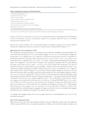
Table 1. Imaging findings suspicious for CCA in PSC patients
Intrahepatic ductal dilatations or DS
Marked dilatation next to strictures
Biliary duct wall thickening
Irregular ductal narrowing with shouldered margins
Intraductal polypoid lesions (> 1 cm)
Focal bile duct thickening with enhancement at MRI
Hyperenhancement in more delayed contrast enhanced phases
Rapid progression of strictures
Focal biliary duct dilatation associated with ipsilateral lobar atrophy
DS: Dominant strictures; MRI: magnetic resonance imaging; ERCP: endoscopic retrograde cholangiopancreatography.
presence of DS is not a synonym of CCA since 50% of patients with PSC will develop focal DS during the
course of the disease , and in a retrospective analysis of 230 patients with PSC, only 19% of biliary
[31]
strictures were caused by CCA .
[32]
Based on the current evidence, the recommended strategy for imaging screening of CCA in PSC patients
without other additional risk factors would be an annual contrast-enhanced MRCP [Figure 1] [20,33] .
Biomarkers for CCA surveillance in PSC
The advantages of using biomarkers for screening are the worldwide availability and reproducibility. The
most widely used biomarker in CCA is carbohydrate antigen 19.9 (CA 19-9). Unfortunately, its diagnostic
performance is not optimal since it is common to find elevated levels in the context of cholestasis. A
preliminary study from the Mayo Clinic reported an AUC of 0.949 (95%CI: 0.895-1.000), with a 78.6%
sensitivity and 98.5% specificity for CA 19-9 level > 129 IU/mL. Unfortunately, all patients had an advanced-
stage CCA at diagnosis . In a later study at the same center including 73 patients with PSC and evaluating
[34]
the same cut-off value (CA19-9 values > 129 IU/L), 37% of the patients did not present CCA after an
[35]
exhaustive study and a median follow-up of 30 months . Similar results were reported in Sweden . In a
[36]
retrospective analysis evaluating 230 patients followed from 2000 to 2006, 23 developed CCA (15 stage I-II
and 8 stages III-IV). By ROC analysis, the cut-off point with the highest diagnostic yield was 20 IU/mL,
which was associated with a sensitivity, specificity, positive predictive value, and negative predictive value of
78%, 67%, 23%, and 96% respectively . Joint use of CA19-9 and CEA has been explored. Although a study
[32]
that included 74 patients with PSC showed a diagnostic yield of 86% , subsequent studies indicate that this
[37]
combination does not offer an acceptable diagnostic yield [36,38] . The combination of serum tumor markers
and imaging is associated with an improvement in sensitivity, including MRI/MRCP plus CA19-9 with a
cut-off value of 20 U/mL (sensitivity, 100%; specificity, 38%; diagnostic average, 89%) and US plus CA19-9
[32]
with a cut-off value of 20 U/mL (sensitivity, 91%; specificity, 62%; diagnostic average, 93%) . Interestingly,
when both tests were negative (CA 19.9 less than 20 UI/mL and lack of evidence of biliary tract
dilatation/bile duct wall thickening by imaging), the negative predictive value was very high which suggests
that physicians can confidently use negative test results to rule out cancer.
According to the available studies, there is not enough evidence for recommending the use of CA 19-9 for
[39]
surveillance purposes .
Role of the endoscopic tests
Biliary strictures are one of the main problems of PSC as DS can be difficult to characterize. The definition
of DS is based on ERCP and is not considered applicable to MRI given the insufficient spatial resolution and