
Page 112 - Read Online
P. 112
Best et al. Hepatoma Res 2020;6:62 I http://dx.doi.org/10.20517/2394-5079.2020.56 Page 3 of 13
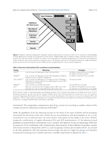
Figure 1. Triangle of hepatocarcinogenesis in metabolic syndrome: hepatocellular carcinoma (HCC) risk in nonalcoholic steatohepatitis
(NASH) significantly increases with progression of liver fibrosis. Independently, type II diabetes mellitus promotes progression from
nonalcoholic fatty liver (NAFL) to NASH, but needs to be recognized as an individual predisposing HCC-risk factor since peripheral
[4]
insulin resistance may promote hepatocarcinogenesis even in the absence of cirrhosis . Microbiome alterations, sedentary lifestyle,
genetic polymorphisms, and obesity represent additional factors aggravating HCC risk in the NASH population
Table 1. Overview of international HCC surveillance recommendations
Society Risk group Procedure
DGVS [15] Liver cirrhosis of all etiologies: chronic HBV and NASH US with or without AFP every 6 months,
US- quality standards required
AASLD [16] Liver cirrhosis of all etiologies; chronic HBV depending on ethnical US with or without AFP every 6 months
background, age, and genetic background
EASL/EORCT [17] Liver cirrhosis of all etiologies at CTP stage A and B or CTP stage C US every 6 months
if listed for oLT; chronic hepatitis B or active hepatitis; chronic HCV
with advanced (F3) fibrosis
APASL [18,19] Liver cirrhosis, chronic HBV and/or HCV US and AFP every 6 months
JSH [20] High risk: Liver cirrhosis of all etiologies; chronic HBV and/or HCV US, AFP, AFP-L3 and DCP every 6 months
Very high risk: Liver cirrhosis with chronic HBV and/or HCV US, AFP, AFP-L3 and DCP every 3-4 months
DGVS: German Society of Gastroenterology and Metabolic Diseases; EASL: European Association for the Study of the Liver; AASLD:
American Association for the Study of Liver Disease; APASL: Asian Pacific Association for the Study of the Liver; JSH: Japanese
Society of Hepatology; HBV: hepatitis B virus; NASH: non-alcoholic Steatohepatits; HCV: hepatitis C virus; US: ultrasound; AFP: alpha
fetoprotein; AFP-L3: Lektin reactive α-Fetoprotein; DCP: Des-gamma-carboxy prothrombin also known as Protein-Induced-by-Vitamin-
K-Absence-or-Antagonist-II (PIVKA II); CTP: Child Turcotte Pugh
determined. The sonographic examinations should be carried out according to quality criteria of the
German Society for Ultrasound in Medicine (DEGUM) .
[15]
While the guidelines of the the American Society for the Study of the Liver (AASLD) and the European
Association for the Study of the Liver (EASL) do not recommend an AFP determination at all, or only
recommend it on an optional basis, the Asian Pacific Association for the Study of the Liver (APASL)
proposed a combination of regular ultrasound (US) and AFP determination. All the aforementioned
guidelines recommend those examinations every 6 months. The Japanese Society for Hepatology (JSH) also
recommends US in combination with complimentary determination of three different biomarkers, namely
AFP, lectin-reactive-α-fetoprotein (AFP-L3), and des-gamma-carboxy-prothrombin (DCP). In contrast
to all other guidelines, JSH discriminates between high risk and very high risk groups and therefore
recommends screening every 6 months and every 3 months, respectively, as depicted in Table 1.