
Page 21 - Read Online
P. 21
Burlone et al. Hepatoma Res 2020;6:3 I http://dx.doi.org/10.20517/2394-5079.2019.37 Page 3 of 10
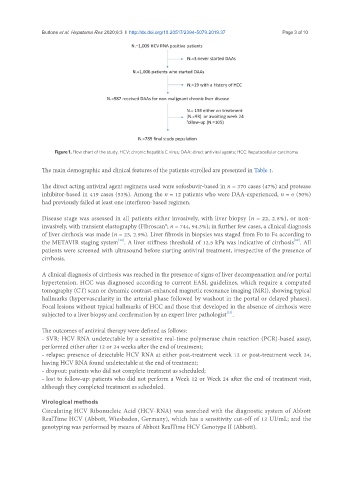
Figure 1. Flow chart of the study. HCV: chronic hepatitis C virus; DAA: direct antiviral agents; HCC: hepatocellular carcinoma
The main demographic and clinical features of the patients enrolled are presented in Table 1.
The direct acting antiviral agent regimens used were sofosbuvir-based in n = 370 cases (47%) and protease
inhibitor-based in 419 cases (53%). Among the n = 12 patients who were DAA-experienced, n = 6 (50%)
had previously failed at least one interferon-based regimen.
Disease stage was assessed in all patients either invasively, with liver biopsy (n = 22, 2.8%), or non-
invasively, with transient elastography (Fibroscan®; n = 744, 94.3%); in further few cases, a clinical diagnosis
of liver cirrhosis was made (n = 23, 2.9%). Liver fibrosis in biopsies was staged from F0 to F4 according to
[12]
[11]
the METAVIR staging system . A liver stiffness threshold of 12.5 kPa was indicative of cirrhosis . All
patients were screened with ultrasound before starting antiviral treatment, irrespective of the presence of
cirrhosis.
A clinical diagnosis of cirrhosis was reached in the presence of signs of liver decompensation and/or portal
hypertension. HCC was diagnosed according to current EASL guidelines, which require a computed
tomography (CT) scan or dynamic contrast-enhanced magnetic resonance imaging (MRI), showing typical
hallmarks (hypervascularity in the arterial phase followed by washout in the portal or delayed phases).
Focal lesions without typical hallmarks of HCC and those that developed in the absence of cirrhosis were
[13]
subjected to a liver biopsy and confirmation by an expert liver pathologist .
The outcomes of antiviral therapy were defined as follows:
- SVR: HCV RNA undetectable by a sensitive real-time polymerase chain reaction (PCR)-based assay,
performed either after 12 or 24 weeks after the end of treatment;
- relapse: presence of detectable HCV RNA at either post-treatment week 12 or post-treatment week 24,
having HCV RNA found undetectable at the end of treatment;
- dropout: patients who did not complete treatment as scheduled;
- lost to follow-up: patients who did not perform a Week 12 or Week 24 after the end of treatment visit,
although they completed treatment as scheduled.
Virological methods
Circulating HCV Ribonucleic Acid (HCV-RNA) was searched with the diagnostic system of Abbott
RealTime HCV (Abbott, Wiesbaden, Germany), which has a sensitivity cut-off of 12 UI/mL; and the
genotyping was performed by means of Abbott RealTime HCV Genotype II (Abbott).