
Page 213 - Read Online
P. 213
Matsushita et al. Hepatoma Res 2018;4:61 I http://dx.doi.org/10.20517/2394-5079.2018.81 Page 7 of 17
A B
C
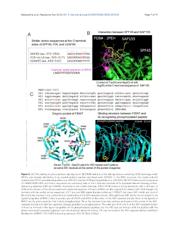
Figure 3. (A) The binding structure between splicing factor 3B (SF3B) and one of the splicing factors containing U2AF homology motif,
SPF45, was already clarified by X-ray crystal analysis (protein data base code: #2PEH). In the 2PEH structure, the crystal unit cell
contains two SPF45 recombinant proteins (a.a. 301-401) and two SF3B partial peptides (a.a. 333-342); (B) SPF45 has an amino sequence
of LNGRYFGGRVVKA and similar sequences are commonly seen at the C-terminal domains of far upstream element-binding protein-
interacting repressor (FIR) and U2AF65. According to the crystal structure, 2PEH, SPF45 makes a strong interaction with a WD part of
SF3B at the domain of the above-mentioned conserved sequence. FIR and U2AF65 are also expected to interact with SF3B through the
domains with the similar amino sequences; (C) F-box and WD repeat domain-containing 7 (FBW7) has many WD motifs and most of
the motifs are involved in the conformational stabilization of the WD-repeated domain. Although those WD motifs are not related to the
ligand recognition of FBW7, there is an extra pair of W425 and D399 at the center of the WD-repeated domain. Most of the ligands of
FBW7 are the amino peptides that include phosphorylated Thr or Ser, because three Arg residues are located at the center of the WD-
repeated domain and hold the negatively charged peptides by phosphorylation. The extra pair of W and D at the WD-repeated domain
will not be involved in the ligand recognition of the phosphorylated peptides, but the WD pair can interact with the peptide with the
above-mentioned conserved sequence from the structural viewpoint. Hence, FIR may be bound to the WD-repeated domain and block
the function of FBW7. FIR: FUBP1-interacting repressor; WD: W (Typ) D (Asp)