Page 210 - Read Online
P. 210
Page 4 of 17                                        Matsushita et al. Hepatoma Res 2018;4:61  I  http://dx.doi.org/10.20517/2394-5079.2018.81


               IUPAC name                                             Structure       His-FIR  His-FIRΔexon2


               1,4a-Dimethyl-2,3,4,4a,9,9a-hexahydro-1H-fluorene-1,9-dicarboxylic acid            3+


               1-(1,4-Dimethyl-1H-pyrazol-3-yl)-2-(4-methoxy-6-methyl-5,6,7,8-         2+
               tetrahydro-[1,3]dioxolo[4,5-g]iso quinolin-5-yl)-ethanone


               1-[2-(1-Ethoxycarbonyl-3-phenyl-propylamino)-propionyl]-pyrrolidine-2-
               carboxylic acid (compound with but-2-enedioic acid)                                3+

               tert-Butyl-{1-(4-methoxy-benzyloxymethyl)-4-[2-methoxy-6-(4-
               methoxy-benzyloxymethyl)-5-methyl-tetr ahydro-pyran-2-yl]-2-methyl-     3+
               butoxy}-diphenyl-silane

               5-Butyl-5-[2-(tert-butyl-diphenyl-silanyloxy)-1-triethylsilanyloxy-ethyl]-  3+     3+
               dihydro-furan-2-one


               3’, 5’, 6’, 7’, 8’, 8’a- hexahydro- 6’- hydroxy- 5’, 8’a- dimethyl- , (5’S, 6’S, 8’aS) -
               Spiro[1, 3- dioxolane- 2, 1’(2’H) - naphthalene] - 5’- acetamide                   2+


               6-iodo-4-methylspiro[3,4-dihydro-1H-quinolin-1-ium-2,1’-cyclohexane]    2+

               3-(2,2-Dimethyl-tetrahydro-pyran-4-yl)-3-[2-(17-hydroxy-10,13-
               dimethyl-1,2,6,7,8,9,10,11,12,13,14,15,16,17-tetradecahydro-cyclopenta[a]  3+
               phenanthren-3-ylideneaminooxy)-acetylamino]-propionic acid

               2-({4-[(2-tert-Butoxycarbonylamino-4-methyl-pentanoylamino)-
               methyl]-cyclohexanecarbonyl}-amino)-4-methylsulfanyl-butyric acid        1+

               6-{2-[3-(2-Methoxy-phenoxy)-2-methyl-4-oxo-4H-chromen-7-yloxy]-
               acetylamino}-hexanoic acid                                              3+


               [2-(2-Benzyloxycarbonylamino-acetylamino)-acetylamino]-acetic acid      3+


               3,7-Bis-furan-2-ylmethylene-bicyclo[3.3.1]nonane-2,6-dione               1+


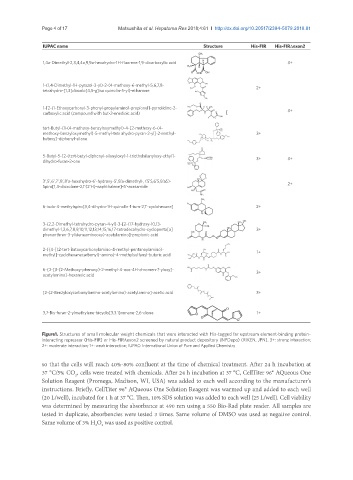
               Figure1. Structures of small molecular weight chemicals that were interacted with His-tagged far upstream element-binding protein-
               interacting repressor (His-FIR) or His-FIRΔexon2 screened by natural product depository (NPDepo) (RIKEN, JPN). 3+: strong interaction;
               2+: moderate interaction; 1+: weak interaction; IUPAC: International Union of Pure and Applied Chemistry


               so that the cells will reach 40%-80% confluent at the time of chemical treatment. After 24 h incubation at
               37 °C/5% CO , cells were treated with chemicals. After 24 h incubation at 37 °C, CellTiter 96® AQueous One
                          2
               Solution Reagent (Promega, Madison, WI, USA) was added to each well according to the manufacturer’s
               instructions. Briefly, CellTiter 96® AQueous One Solution Reagent was warmed up and added to each well
               (20 L/well), incubated for 1 h at 37 °C. Then, 10% SDS solution was added to each well (25 L/well). Cell viability
               was determined by measuring the absorbance at 490 nm using a 550 Bio-Rad plate reader. All samples are
               tested in duplicate, absorbencies were tested 3 times. Same volume of DMSO was used as negative control.
               Same volume of 3% H O  was used as positive control.
                                   2
                                 2

