
Page 167 - Read Online
P. 167
Page 4 of 11 Tamai. Hepatoma Res 2018;4:75 I http://dx.doi.org/10.20517/2394-5079.2018.98
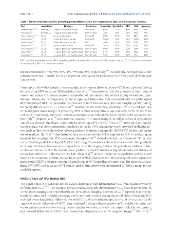
Table 1. Studies with ultrasound for predicting poorly differentiation, non-single nodular type, or microvascular invasion
Ref. Modalities Findings Prediction Sensitivity Specificity PPV NPV Accuracy
Moribata et al. [10] B-mode US Irregular or unclear margin Poorly diff. 89% 67% 19% 99% 69%
Nuta et al. [21] B-mode US Irregular or unclear margin Non-SN type 72% 85% 96% 39% 74%
Sugimoto et al. [11] CEUS Dead wood pattern Poorly diff. 80% 96% 86% 94% 92%
Tanaka et al. [12] CEUS Grade 4 (hypo-irregular) Poorly diff. 100% 91% 50% 100% 92%
Tanaka et al. [12] CEUS Grade 4 (hypo-irregular) MVI 40% 92% 67% 80% 78%
Feng et al. [17] CEUS Washout time < 40 s Poorly diff. 24% 97% 65% 61% 69%
Hatanaka et al. [18] CEUS Irregular defect on Kupffer phase Non-SN type 80% 96% 92% 89% 90%
Tada et al. [19] CEUS Irregular defect on Kupffer phase Non-SN type 87% 93% 91% 84% 95%
Nuta et al. [21] CEUS Irregular defect on Kupffer phase Non-SN type 93% 85% 97% 73% 92%
PPV: positive predictive value; NPV: negative predictive value; US: ultrasound; SN: single nodular; CEUS: contrast enhanced
ultrasonography; MVI: microvascular invasion
[26]
tumor enhancement were 75%, 90%, 48%, 97% and 88%, respectively . Accordingly, heterogenous tumor
enhancement even in small HCC is an important observation for predicting HCC with poorly differentiated
components.
Some reports show that irregular tumor margin in the venous phase of contrast CT is an important finding
[27]
for predicting MVI or tumor differentiation. Lee et al. demonstrated that the presence of intra-tumoral
vessels and aneurysms, tumor necrosis, attenuation of pre-contrast, the relative timing of washout, intra-
tumoral attenuation heterogeneity, tumor margin, and tumor size were correlated with the pathological
differentiation of HCC. In particular, the presence of intra-tumoral aneurysm was a highly specific finding
[28]
for poorly differentiated HCC. Chou et al. showed that the sensitivity, specificity, PPV, NPV, and accuracy
of the irregular tumor margin in predicting MVI in their retrospective study were 66%, 86.5%, 82.5%, 72.6%
and 76.5%, respectively, and those in their prospective study were 81.7%, 88.1%, 90.7%, 77.1% and 84.3%, re-
[29]
[30]
spectively . Reginelli et al. indicated that irregularity in tumor margins, as well as defects of peritumoral
[31]
capsule are the most significant characteristics predicting MVI in HCC. Wu et al. reported that irregular
tumor margin was alone independent predictive factor for MVI among previously proposed predicting fac-
tors such as fluorine-18 fluorodeoxyglucose-positron emission tomography (FDG-PET) results and serum
[32]
tumor markers. Hu et al. demonstrated in a meta-analysis that CT is superior to MRI in evaluating an
[33]
irregular tumor margin for MVI assessment. Banerjee et al. showed new features of contrast CT that can
also accurately predict histological MVI in HCC surgical candidates. These features include: the positivity
of radiogemic venous invasion consisting of three separate imaging features; the persistence of discrete arte-
rial tumor enhancement in the venous phase; partial or complete absence of hypodense halo; and absence of
[34]
tumor-liver difference in the absence of a halo. Zhao et al. demonstrated that the predictive scoring model
based on intra-tumoral arteries, non-nodular type of HCC, and absence of the radiological tumor capsule on
preoperative CECT is of great value in the prediction of MVI regardless of tumor size. The sensitivity, speci-
ficity, PPV, NPV, and accuracy of CT studies cited in this review were summarized in Table 2 except for not
available reports.
PREDICTION OF MVI USING MRI
The signal intensity of MRI can also be used to distinguish well differentiated HCC from moderately/poorly
differentiated HCC [35-37] . For example, typical moderately/poorly differentiated HCC show hypointensity on
[38]
T1-weighted imaging and hyperintensity on T2-weighted imaging. Enomoto et al. reported that hypoin-
tensity of tumor on T1-weighted imaging and tumor stain washout during the portal phase of dynamic MRI
reflected poorer histological differentiation of HCCs, and the sensitivity, specificity, and the accuracy for di-
agnosis of poorly differentiated HCC using combined findings of hypointensity on T1-weighted imaging and
tumor enhancement washout during the portal phase were 88%, 67% and 71%, respectively. On the contrary,
[39]
[38]
most of well-differentiated HCCs (83%) showed non-hypointensity on T1-weighted image . Min et al.