
Page 90 - Read Online
P. 90
Loria et al. Hepatoma Res 2018;4:59 I http://dx.doi.org/10.20517/2394-5079.2018.75 Page 7 of 12
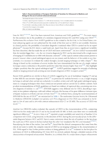
Table 3. Recommendations of European Federation of Societies for Ultrasound in Medicine and
Biology for the use of contrast-enhanced ultrasoud
The characterization of the nodules
To make a rapid diagnosis (however, CT or MR remain necessary, if not contraindicated, for the stadiation
When CT and MR are inconclusive especially in nodules that can’t be submitted to biopsy
To contribute to selecting a nodule when they are many or have different contrast patterns
To monitor the changes in the nodule
After an inconclusive histology
lines for HCC [22,23,65,66] , but it has been removed from American and EASL guidelines [48,53] . The main reason
for this exclusion lies in the possibility of a mistaken diagnosis between ICC and HCC using only CEUS [67,68] .
Furthermore this exclusion from AASLD guidelines is also related to the fact that, in the United States, con-
trast enhancing agents are not authorized for the study of the liver and so CEUS is not available. However,
in clinical practice, the probability of mistaken diagnosis is minimal when CEUS is carried out by an expert
[69]
physician , because the ICC shows a rapid wash-out. Apart from this, in recent years a significant variability
[69]
has been described, that has made the use of CEUS still more controversial . In 2010 AASLD recommended
that, for nodules bigger than 1 cm, the non invasive diagnosis for HCC can be determined with a single means
[53]
of imaging (CT multidetector or MRI with dynamic contrast) , if the typical contrast enhancement pattern is
present; however when typical radiological aspects are not present and the behavior of the nodule is not char-
acteristic, it is necessary to evaluate the nodule through a second imaging technique or with a biopsy . This
[53]
change is based on the conclusion of several studies that have demonstrated that the use of a single contrast
technique causes a reduction in the positive predictive value that remains higher than 90% [42,59] , they highlight
a higher specificity than the typical radiological sigh [41,70] . AASLD guidelines suggest the necessity of adhering
closely to imaging protocol and carrying out non invasive diagnosis of HCC in expert centers [2,53] .
Recent EASL guidelines are similar to those of AASLD, suggesting the use of multiphase imaging CT and up
to date MRI for non invasive diagnosis of HCC ; in particular for nodules between 1-2 cm, a single imaging
[48]
technique is advised when carried out exclusively in excellent centers and with high grade radiological equip-
ment or 2 imaging techniques when these criteria are not present and are carried out in inferior contexts. Such
prudent recommendations of EASL guidelines are based on evidence of equivocal data concerning non inva-
sive diagnosis of nodules 1-2 cm [22,48,53] . EFSUMB suggests a very different role for CEUS, describing it sepa-
rately in two patients subgroups, with and without cirrhosis; this because of the great difference between types
of hepatic nodules in cirrhotic and non cirrhotic livers [22-23] . In cirrhotic livers, among the recommendations of
[23]
EFSUMB for the use of CEUS are summarized in Table 3. The multicenter German Society for Ultrasound
in Medicine (DEGUM) included 1349 patients with FLLs diagnosed on US; CEUS was compared to the bi-
opsy in 75% of cases and in 25% with contrast enhancement (CE) CT or CE-MRI. The accuracy of CEUS was
90.3% [71-75] .
Another two DEGUM studies evaluated the capacity of CEUS in the characterization of FLL, comparing
CEUS in the first study with CE-CT and in the second with CE-MR. In both cases there were no statistically
[78]
significant differences [75-77] . In 2012, Goto et al. reported a major sensibility and sensitivity of baseline US in
comparison with CEUS, using Sonazoid, in the detection of HCC during the post-vascular phase. In the differ-
ential diagnosis between HCC and ICC there is some controversy about the role of washout: in the late phase
the wash-out of HCC seems to be less marked than the other liver neoplasms like ICC and metastasis [23,38,69,79] .
[80]
Reanalyzing the data of the studies, Guo and Xu , found that the clinical consequences that come from this
risk do not seem to justify the complete removal of CEUS as an imaging technique in the characterization of
[81]
FLL. With regard to this, further positive evidence is being gathered: Li et al. evaluated in the first place the
usefulness of CEUS in differentiating ICC from HCC in cirrhotic patients through a detailed analysis of the
characteristics of temporal enhancement. Therefore, in a cirrhotic liver if a nodule shows a hyper-enhancement