
Page 10 - Read Online
P. 10
Page 4 of 10 Wang et al. Hepatoma Res 2018;4:14 I http://dx.doi.org/10.20517/2394-5079.2018.16
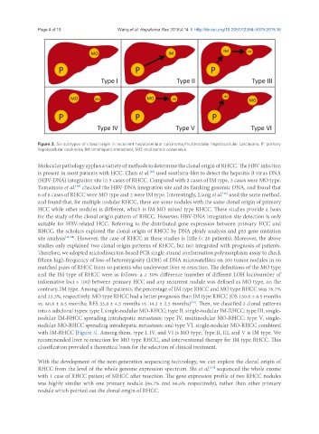
Figure 3. Six subtypes of clonal origin in recurrent hepatocellular carcinoma/multinodular hepatocellular carcinoma. P: primary
hepatocellular carcinoma; IM: intrahepatic metastasis; MO: multicentric occurrence
Molecular pathology applies a variety of methods to determine the clonal origin of RHCC. The HBV infection
is present in most patients with HCC. Chen et al. used southern-blot to detect the hepatitis B virus DNA
[49]
(HBV-DNA) integration site in 5 cases of RHCC. Compared with 2 cases of IM type, 3 cases were MO type.
Yamamoto et al. checked the HBV-DNA integration site and its flanking genomic DNA, and found that
[50]
6 of 8 cases of RHCC were MO type and 2 were IM type. Interestingly, Liang et al. used the same method,
[51]
and found that, for multiple nodular RHCC, there are some nodules with the same clonal origin of primary
HCC while other nodules is different, which is IM-MO mixed type RHCC. These studies provide a basis
for the study of the clonal origin pattern of RHCC. However, HBV-DNA integration site detection is only
suitable for HBV-related HCC. Referring to the distributed gene expression between primary HCC and
RHCC, the scholars explored the clonal origin of RHCC by DNA ploidy analysis and p53 gene mutation
site analysis [52-54] . However, the case of RHCC in these studies is little (< 20 patients). Moreover, the above
studies only explained two clonal origin patterns of RHCC, but not integrated with prognosis of patients.
Therefore, we adopted microdissecton-based PCR single-strand conformation polymorphism assay to check
fifteen high-frequency of loss of heterozygosity (LOH) of DNA microsatellites on 100 tumor nodules in 60
matched pairs of RHCC from 40 patients who underwent liver re-resection. The definitions of the MO type
and the IM type of RHCC were as follows: a ≥ 30% difference (number of different LOH loci/number of
informative loci × 100) between primary HCC and any recurrent nodule was defined as MO type, on the
contrary, IM type. Among all the patients, the percentage of IM type RHCC and MO type RHCC was 76.7%
and 23.3%, respectively. MO type RHCC had a better prognosis than IM type RHCC (OS 130.8 ± 8.5 months
vs. 80.8 ± 8.5 months; RFS 33.8 ± 4.5 months vs. 14.2 ± 2.5 months) . Then, we classified 2 clonal patterns
[33]
into 6 subclonal types: type I, single-nodular MO-RHCC; type II, single-nodular IM-RHCC; type III, single-
nodular IM-RHCC spreading intrahepatic metastasis; type IV, multinodular MO-RHCC; type V, single-
nodular MO-RHCC spreading intrahepatic metastasis; and type VI, single-nodular MO-RHCC combined
with IM-RHCC [Figure 3]. Among them, type I, IV, and VI is MO type; Type II, III, and V is IM type. We
recommended liver re-resection for MO type RHCC, and interventional therapy for IM type RHCC. This
classification provided a theoretical basis for the selection of clinical treatment.
With the development of the next-generation sequencing technology, we can explore the clonal origin of
RHCC from the level of the whole genome expression spectrum. Shi et al. sequenced the whole exome
[55]
with 1 case of RHCC patient of MHCC after resection. The gene expression profile of two RHCC nodules
was highly similar with one primary nodule (86.7% and 86.6% respectively), rather than other primary
nodule which pointed out the clonal origin of RHCC.