
Page 15 - Read Online
P. 15
Fabbrizi et al. Cancer Drug Resist 2020;3:775-90 I http://dx.doi.org/10.20517/cdr.2020.49 Page 777
molecules. Low linear energy transfer (LET) radiation, including X-rays and g-rays, deposits a relatively
small amount of energy over a short distance, whereas high LET radiation, including charged particles, will
deposit a large amount of energy within the same short distance [14,15] . Consequently, high LET radiation
will display an enhanced biological effect. This is in part mediated by increases in the formation of DNA
DSBs and CDD that represent a greater challenge to the cellular DNA repair machinery, whereas the
majority of DNA lesions generated by low-LET radiation are DNA base lesions and SSBs that are repaired
relatively more efficiently. Particle beam therapy (e.g., proton or carbon ion beams) displays another
distinct advantage in that the radiation dose can be delivered directly to the cancer cells with limited
exposure of the surrounding healthy tissue. This is due to the deposition of maximum energy in depth at a
well-defined region, the Bragg peak, which in turn will lead to less RT-related side effects when compared
[16]
to conventional RT using low-LET photons . However, particle beam therapy exhibits increases in LET
at and around the Bragg peak, which can generate a different DNA damage spectra that contributes to an
increased biological response. As a consequence, there is still a significant amount of biological uncertainty
when utilising this therapeutic approach [17,18] . Nevertheless, the main goal of RT is to either cause sufficient
cellular DNA damage or enhance the persistence of the damage to greatly exceed the cells capacity for
repair, thus promoting cell death.
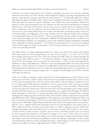
The cellular DDR is an elegant signaling network able to detect and repair DNA damage, with different
DNA repair pathways activated depending on the type of lesion [Figure 1]. At least 90%-95% of the
DNA damage induced by low-LET RT is DNA base damage and SSBs, which are repaired through the
base excision repair (BER) pathway [19,20] . The DNA base damage is recognized and excised by damage-
specific DNA glycosylases, which along with AP endonuclease-1 (APE1), incise the phosphodiester
backbone to create an SSB flanked by 3’-OH and 5’-deoxyribosephosphate (dRP) ends. Poly (ADP-ribose)
polymerase-1 (PARP-1) binds to the SSB due to its high affinity for the lesion and protects the DNA ends.
DNA polymerase b (Pol b) then excises the 5’-dRP ends and simultaneously inserts a new nucleotide. This
is followed by X-ray cross-complementing protein 1 in complex with DNA ligase IIIa (XRCC1-Lig IIIa)
which ligate the SSB ends.
In the case of DSBs, the damage is firstly recognized by the ataxia telangiectasia mutated (ATM) or ataxia
telangiectasia and Rad-3 related (ATR) protein kinases that stimulate phosphorylation of the histone
H2AX variant (forming gH2AX). Cells then activate either NHEJ or HR depending on whether the cell is
in G /G phase where NHEJ pathway is employed, or in S/G phases where a sister chromatid is available
2
0
1
[12]
to enable HR. NHEJ can be further divided into two sub-pathways . Canonical-NHEJ (c-NHEJ) utilises
the Ku70/80 heterodimer that bind the DSB ends, followed by the DNA-dependent protein kinase catalytic
subunit (DNA-Pkcs) and X-ray cross-complementing protein 4 in complex with DNA ligase IV (XRCC4/
LigIV) that ligate the DSB ends. In contrast, alternative-NHEJ (a-NHEJ) is initiated by the C-terminal
binding protein-interacting protein (CtIP) and MRE11/RAD50/NBS1 (MRN) complex that resect the DNA
ends, followed by PARP-1 binding, gap filling by a DNA polymerase, and ultimately ligation by XRCC1-Lig
IIIa (or alternatively DNA ligase I). HR is an error-free DSB repair mechanism, and similar to a-NHEJ, is
initiated by the CtIP-MRN complex that promotes DNA end resection and requires breast cancer protein 1
(BRCA1). The 3’-single stranded DNA generated is then bound and stabilized by replication protein A
(RPA), replaced with the recombination protein RAD51 in a BRCA2-dependent manner, which then
undergoes homology search and strand invasion into the sister chromatid. DNA synthesis in the presence
of RAD52/RAD54 results in branch migration and formation of Holliday junctions that are resolved
by DNA resolvases that ultimately restore genome integrity. In the case of CDD, which can include a
mixture of DNA lesions within a short distance on the DNA molecule, it is likely that the repair of these
sites requires more than one DNA repair pathway to resolve the damage. Interestingly, we have recently
demonstrated that high-LET proton irradiation generates a high frequency of CDD sites consisting of
[21]
abasic sites and SSBs, which have a high dependency on PARP-1 for repair .