
Page 24 - Read Online
P. 24
Brettrager et al. Cancer Drug Resist 2019;2:1153-63 I http://dx.doi.org/10.20517/cdr.2019.91 Page 1157
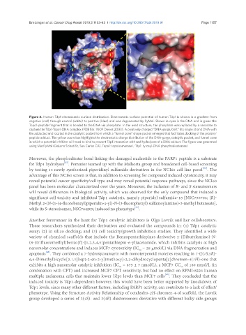
Figure 2. Human Tdp1 electrostatic surface distribution. Electrostatic surface potential of human Tdp1 is shown in a gradient from
negative (red) through neutral (white) to positive (blue) and was degenerated by PyMol. Shown in cyan is the DNA and in green the
Topo1-peptide fragment that is bonded to the DNA via phosphate. In the used structure, the phosphate was replaced by a vanadate to
capture the Tdp1-Topo1-DNA complex (PDB file: 1NOP Davies 2003). A positively charged “DNA-gorge/cleft” fits single strand DNA with
the adducted end located in the catalytic pocket from which a “funnel cone” shape pocket emerges that facilitates docking of the protein/
peptide adduct. The yellow zoom box highlights the electrostatic charge distribution of the DNA-gorge, catalytic pocket, and funnel cone
in which a potential inhibitor will need to bind to prevent Tdp1 interaction with and hydrolyzes of a DNA-adduct. The figure was generated
using MacPyMol (DeLano Scientific, San Carlos CA). Topo1: topoisomerase I; Tdp1: tyrosyl-DNA phosphodiesterase I
Moreover, the phosphodiester bond linking the damaged nucleotide to the PARP1 peptide is a substrate
[23]
for Tdp1 hydrolyses . Pommier teamed up with the Malhorta group and broadened cell-based screening
[61]
by testing 15 newly synthesized piperidinyl sulfamide derivatives in the NCI60 cell line panel . The
advantage of this NCI60 screen is that, in addition to screening for compound induced cytotoxicity, it may
reveal potential cancer specificity/cell type and may reveal potential response pathways, since the NCI60
panel has been molecular characterized over the years. Moreover, the inclusion of R- and S-stereoisomers
will reveal differences in biological activity, which was observed for the only compound that induced a
significant cell toxicity and inhibited Tdp1 catalysis, namely piperidyl sulfamide-18 [NSC750706; (R)-
Methyl 2-(N-(1-(4-fluorobenzyl)piperidin-4-yl)-N-(3-fluorophenyl) sulfamoylamino)-3-methyl butanoate],
[61]
while its S-stereoisomer, NSC764209, induced no phenotype .
Another forerunner in the hunt for Tdp1 catalytic inhibitors is Olga Lavrik and her collaborators.
These researchers synthesized their derivatives and evaluated the compounds in: (1) Tdp1 catalytic
assay; (2) in silico docking; and (3) cell toxicity/growth inhibition studies. They identified a wide
variety of chemical scaffolds that include the Benzopentathiepines derivative 2-(Dibutylamino)-N-
(8-(trifluoromethyl)benzo[f]-[1,2,3,4,5]pentathiepin-6-yl)acetamide, which inhibits catalysis at high
nanomolar concentrations and induces MCF7 cytotoxicity (IC ~ 28 μmol/L) via DNA fragmentation and
50
[62]
apoptosis . They combined a 7-hydroxycoumarin with monoterpenoid moieties resulting in 7-(((1S,5R)-
6,6-Dimethylbicyclo[3.1.1]hept-2-en-2-yl)methoxy)-2,3-dihydrocyclopenta[c]chromen-4(1H)-one that
exhibits a high nanomolar catalytic inhibition (IC = 675 ± 7 nmol/L), a MCF7 CC of 180 nmol/L (in
50
50
combination with CPT) and increased MCF7 CPT sensitivity, but had no effect on RPMI-8226 human
[63]
multiple melanoma cells that maintain lower Tdp1 levels than MCF7 cells . They concluded that the
induced toxicity is Tdp1-dependent; however, this would have been better supported by knockdown of
Tdp1 levels, since many other different factors, including PARP1 activity, can contribute to a lack of effect/
phenotype. Using the Structure-Activity Relationship of octahydro-2H-chromen-4-ol scaffold, the Lavrik
group developed a series of 3(4S)- and 3(4R)-diastereomers derivative with different bulky side-groups