
Page 23 - Read Online
P. 23
Page 1156 Brettrager et al. Cancer Drug Resist 2019;2:1153-63 I http://dx.doi.org/10.20517/cdr.2019.91
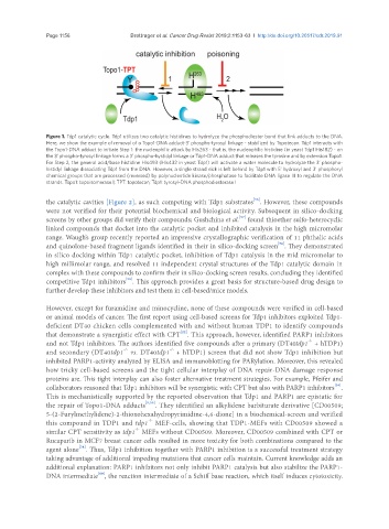
Figure 1. Tdp1 catalytic cycle. Tdp1 utilizes two catalytic histidines to hydrolyze the phosphodiester bond that link adducts to the DNA.
Here, we show the example of removal of a Topo1-DNA adduct-3’ phospho-tyrosyl linkage - stabilized by Topotecan. Tdp1 interacts with
the Topo1-DNA adduct to initiate Step 1: the nucleophilic attack by His263 - that is, the nucleophilic histidine (in yeast Tdp1 His182) - on
the 3’ phospho-tyrosyl linkage forms a 3’ phospho-hystidyl linkage or Tdp1-DNA adduct that releases the tyrosine and by extension Topo1.
For Step 2, the general acid/base histidine His493 (His432 in yeast Tdp1) will activate a water molecule to hydrolyze the 3’ phospho-
histidyl linkage dissociating Tdp1 from the DNA. However, a single strand nick is left behind by Tdp1 with 5’ hydroxyl and 3’ phosphoryl
chemical groups that are processed (reversed) by polynucleotide kinase/phosphatase to facilitate DNA ligase III to regulate the DNA
strands. Topo1: topoisomerase I; TPT: topotecan; Tdp1: tyrosyl-DNA phosphodiesterase I
[56]
the catalytic cavities [Figure 2], as such competing with Tdp1 substrates . However, these compounds
were not verified for their potential biochemical and biological activity. Subsequent in silico-docking
[57]
screens by other groups did verify their compounds; Gushchina et al. found thioether sulfo-heterocyclic
linked compounds that docket into the catalytic pocket and inhibited catalysis in the high micromolar
range. Waugh’s group recently reported an impressive crystallographic verification of 11 phthalic acids
[58]
and quinolone-based fragment ligands identified in their in silico-docking screen . They demonstrated
in silico docking within Tdp1 catalytic pocket, inhibition of Tdp1 catalysis in the mid micromolar to
high millimolar range, and resolved 11 independent crystal structures of the Tdp1 catalytic domain in
complex with these compounds to confirm their in silico-docking screen results, concluding they identified
[58]
competitive Tdp1 inhibitors . This approach provides a great basis for structure-based drug design to
further develop these inhibitors and test them in cell-based/mice models.
However, except for furamidine and minocycline, none of these compounds were verified in cell-based
or animal models of cancer. The first report using cell-based screens for Tdp1 inhibitors exploited Tdp1-
deficient DT40 chicken cells complemented with and without human TDP1 to identify compounds
that demonstrate a synergistic effect with CPT . This approach, however, identified PARP1 inhibitors
[22]
-/-
and not Tdp1 inhibitors. The authors identified five compounds after a primary (DT40tdp1 + hTDP1)
-/-
and secondary (DT40tdp1 vs. DT40tdp1 + hTDP1) screen that did not show Tdp1 inhibition but
-/-
inhibited PARP1-activity analyzed by ELISA and immunoblotting for PARylation. Moreover, this revealed
how tricky cell-based screens and the tight cellular interplay of DNA repair-DNA damage response
proteins are. This tight interplay can also foster alternative treatment strategies. For example, Pfeifer and
collaborators reasoned that Tdp1 inhibitors will be synergistic with CPT but also with PARP1 inhibitors .
[34]
This is mechanistically supported by the reported observation that Tdp1 and PARP1 are epistatic for
the repair of Topo1-DNA adducts [9,59] . They identified an alkylidene barbiturate derivative [CD00509;
5-(2-Furylmethylidene)-2-thioxohexahydropyrimidine-4,6-dione] in a biochemical-screen and verified
-/-
this compound in TDP1 and tdp1 MEF-cells, showing that TDP1-MEFs with CD00509 showed a
-/-
similar CPT sensitivity as tdp1 MEFs without CD00509. Moreover, CD00509 combined with CPT or
Rucaparib in MCF7 breast cancer cells resulted in more toxicity for both combinations compared to the
[34]
agent alone . Thus, Tdp1 inhibition together with PARP1 inhibition is a successful treatment strategy
taking advantage of additional impeding mutations that cancer cells maintain. Current knowledge adds an
additional explanation: PARP1 inhibitors not only inhibit PARP1 catalysis but also stabilize the PARP1-
[60]
DNA intermediate , the reaction intermediate of a Schiff base reaction, which itself induces cytotoxicity.