Page 167 - Read Online
P. 167
Belizario et al. Cancer Drug Resist 2019;2:527-38  I  http://dx.doi.org/10.20517/cdr.2018.009                                            Page 531


                                                                       P


                                                                      P


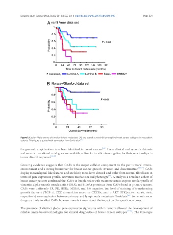
               Figure 1. Kaplan-Meier curves of time for distant metastasis (A) and overall survival (B) among five breast cancer subtypes in two patient
               cohorts. This figure is quoted with permission from Sorlie et al. [33]

               the genomic amplification have been identified in breast cancers . These clinical and genomic datasets
                                                                       [42]
               and somatic mutational catalogues are available online for in silico investigation for their relationships to
               tumor clinical responses [42,43] .


               Growing evidence suggests that CAFs is the major cellular component in the peritumoral micro-
               environment and a strong biomarker for breast cancer growth invasion and dissemination [20,24] . CAFs
               display mesenchymal-like features and are likely mesoderm derived and differ from normal fibroblasts in
                                                                            [44]
               terms of gene expression profile, activation mechanism and phenotype . A study in a Brazilian cohort of
               breast cancer patients confirmed that CAFs in lymph nodes with macrometastasis express similar profile of
               vimentin, alpha-smooth muscle actin (-SMA), and S100A4 protein as those CAFs found in primary tumors.
               CAFs were uniformly ER, PR, HER2, MKI67, and P53 negative, but level of staining of transforming
               growth factor-1 (TGF-1), CXC chemokine receptor CXCR4, and p-AKT STK(62.3%, 52.4%, 65%,
                                                                                         [44]
               respectively) were equivalent between primary and lymph node metastasis fibroblasts . Some anticancer
               drugs are likely to affect CAFs, however none is known about the impact on therapeutic outcomes.

               The presence of distinct global gene-expression signatures within tumors allowed the development of
               reliable omics-based technologies for clinical diagnostics of breast cancer subtypes [33-36] . The Oncotype

