


Page 34 - Read Online
P. 34
Talwar et al. Art Int Surg. 2025;5:418-24  https://dx.doi.org/10.20517/ais.2025.22                                                         Page 422

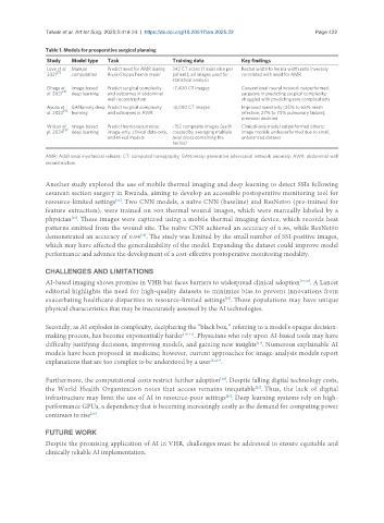
               Table 1. Models for preoperative surgical planning
                Study  Model type  Task               Training data         Key findings
                Love et al.  Manual   Predict need for AMR during   342 CT scans (1 axial slice per   Rectus width to hernia width ratio inversely
                   [5]
                2021   computation  Rives-Stoppa hernia repair  patient); all images used for   correlated with need for AMR
                                                      statistical analysis
                Elhage et   Image-based   Predict surgical complexity   ~7,400 CT images  Conventional neural network outperformed
                    [6]
                al. 2021  deep learning  and outcomes in abdominal          surgeons in predicting surgical complexity;
                                  wall reconstruction                       struggled with predicting rare complications
                Ayuso et   GANomaly deep  Predict surgical complexity   ~8,000 CT images  Improved sensitivity (25% to 68% mesh
                al. 2023 [9]  learning  and outcomes in AWR                 infection, 27% to 73% pulmonary failure);
                                                                            precision declined
                Wilson et  Image-based   Predict hernia recurrence;   ~152 composite images (each   Clinical-only model outperformed others;
                     [10
                al. 2024  deep learning  image-only, clinical data-only,  created by averaging multiple   image models underperformed due to small,
                ]
                                  and mixed models    axial slices containing the   unbalanced dataset
                                                      hernia)
               AMR: Additional myofascial release; CT: computed tomography; GANomaly: generative adversarial network anomaly; AWR: abdominal wall
               reconstruction.


               Another study explored the use of mobile thermal imaging and deep learning to detect SSIs following
               cesarean section surgery in Rwanda, aiming to develop an accessible postoperative monitoring tool for
               resource-limited settings . Two CNN models, a naïve CNN (baseline) and ResNet50 (pre-trained for
                                     [16]
               feature extraction), were trained on 500 thermal wound images, which were manually labeled by a
                       [16]
               physician . These images were captured using a mobile thermal imaging device, which records heat
               patterns emitted from the wound site. The naïve CNN achieved an accuracy of 0.86, while ResNet50
               demonstrated an accuracy of 0.94 . The study was limited by the small number of SSI-positive images,
                                            [16]
               which may have affected the generalizability of the model. Expanding the dataset could improve model
               performance and advance the development of a cost-effective postoperative monitoring modality.


               CHALLENGES AND LIMITATIONS
               AI-based imaging shows promise in VHR but faces barriers to widespread clinical adoption [17,18] . A Lancet
               editorial highlights the need for high-quality datasets to minimize bias to prevent innovations from
                                                                       [19]
               exacerbating healthcare disparities in resource-limited settings . These populations may have unique
               physical characteristics that may be inaccurately assessed by the AI technologies.

               Secondly, as AI explodes in complexity, deciphering the “black box,” referring to a model’s opaque decision-
               making process, has become exponentially harder [19-21] . Physicians who rely upon AI-based tools may have
               difficulty justifying decisions, improving models, and gaining new insights . Numerous explainable AI
                                                                                [21]
               models have been proposed in medicine; however, current approaches for image-analysis models report
               explanations that are too complex to be understood by a user [22,23] .


               Furthermore, the computational costs restrict further adoption . Despite falling digital technology costs,
                                                                     [24]
               the World Health Organization notes that access remains inequitable . Thus, the lack of digital
                                                                                [25]
               infrastructure may limit the use of AI in resource-poor settings . Deep learning systems rely on high-
                                                                       [25]
               performance GPUs, a dependency that is becoming increasingly costly as the demand for computing power
               continues to rise .
                             [26]
               FUTURE WORK
               Despite the promising application of AI in VHR, challenges must be addressed to ensure equitable and
               clinically reliable AI implementation.

