
Page 17 - Read Online
P. 17
Dababneh et al. Art Int Surg 2024;4:214-32 https://dx.doi.org/10.20517/ais.2024.50 Page 222
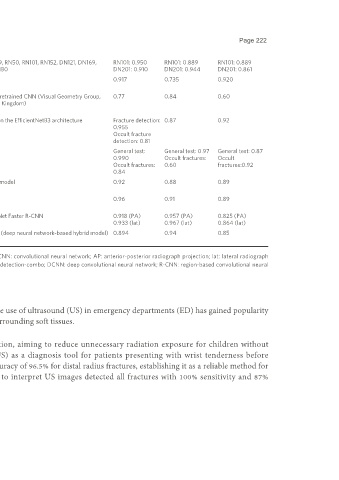
Tung et al. Scaphoid 356 VGG16, VGG19, RN50, RN101, RN152, DN121, DN169, RN101: 0.950 RN101: 0.889 RN101: 0.889
[44]
(2021) DN201, Inv, ENB0 DN201: 0.910 DN201: 0.944 DN201: 0.861
Yang et al. Scaphoid 361 ResNet 0.917 0.735 0.920
[45]
(2022)
Langerhuizen Scaphoid 300 (scaphoid series) Open source pretrained CNN (Visual Geometry Group, 0.77 0.84 0.60
[47]
et al. (2020) Oxford, United Kingdom)
Yoon et al. Scaphoid 11,838 (PA, scaphoid view) DCNN based on the EfficientNetB3 architecture Fracture detection: 0.87 0.92
[48]
(2021) 0.955
Occult fracture
detection: 0.81
Raisuddin et al. Distal radius 4,497 (AP, lat) DeepWrist General test: General test: 0.97 General test: 0.87
[49]
(2021) 0.990 Occult fractures: Occult
Occult fractures: 0.60 fractures:0.92
0.84
Zech et al. Distal radius 395 (AP) Faster R-CNN model 0.92 0.88 0.89
[51]*
(2023)
Ilie et al. Finger, hand, wrist, forearm, elbow, 58,846 Faster R-CNN 0.96 0.91 0.89
[53]
(2023) humerus, shoulder, clavicule
Watanabe Distal radius or distal ulna 7,356 (PA, lat) Inception-ResNet Faster R-CNN 0.918 (PA) 0.957 (PA) 0.825 (PA)
[54]
et al. (2019) 0.933 (lat) 0.967 (lat) 0.864 (lat)
Orji et al. Finger 8,170 ComDNet-512 (deep neural network-based hybrid model) 0.894 0.94 0.85
[55]
(2022)
*
Pediatric studies. ML: Machine learning; AUC: area under the receiver operator characteristic curve; CNN: convolutional neural network; AP: anterior-posterior radiograph projection; lat: lateral radiograph
projection; AI: artificial intelligence; PA: posterior-anterior radiograph projection; WFD-C: wrist fracture detection-combo; DCNN: deep convolutional neural network; R-CNN: region-based convolutional neural
network.
AI as an adjuvant in ultrasound fracture detection
While X-rays remain the gold standard for diagnosing distal radius fractures, the use of ultrasound (US) in emergency departments (ED) has gained popularity
due to its accessibility, minimal training requirements, and capacity to assess surrounding soft tissues.
Zhang et al. were the first to explore the potential of US for fracture detection, aiming to reduce unnecessary radiation exposure for children without
fractures . In their study, they used a three-dimensional ultrasound (3DUS) as a diagnosis tool for patients presenting with wrist tenderness before
[56]
undergoing X-rays. The findings demonstrated that 3DUS had a diagnostic accuracy of 96.5% for distal radius fractures, establishing it as a reliable method for
fracture detection in a pediatric setting. Moreover, the CNN model trained to interpret US images detected all fractures with 100% sensitivity and 87%
specificity, matching the sensitivity of the pediatric MSK radiologist.