
Page 7 - Read Online
P. 7
Gillion et al. Rare Dis Orphan Drugs J 2023;2:11 https://dx.doi.org/10.20517/rdodj.2022.23 Page 3 of 5
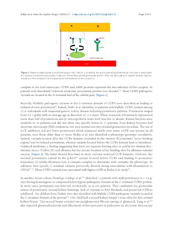
Figure 1. Albumin reabsorption in proximal tubular cells. Cubilin is located in the apical part of proximal tubular cells and is responsible
for receptor-mediated endocytosis of albumin filtered through the glomerular barrier. After the decoupling of cubulin-albumin ligation,
albumin is then released from the basolateral cell surface into the circulation.
complex in the ileal enterocyte. CUBN and AMN proteins represent the two subunits of this receptor. In
patients with Imerslund-Gräsbeck syndrome, proteinuria persists over decades . Most CUBN pathogenic
[7,8]
variants are located in the N-terminal half of the cubilin gene [Figure 2].
Recently, bilallelic pathogenic variants in the C-terminal domain of CUBN were described as leading to
isolated chronic proteinuria . Indeed, Bedin et al. identified 39 patients with biallelic CUBN variants among
[4]
2216 individuals with suspected genetic kidney disease including proteinuric patients. Proteinuria ranged
from 0,5-3 g/day with an average age at discovery of 10.9 years. When measured, albuminuria represented
more than half of proteinuria and β2-microglobulin urine level was low or absent. Kidney biopsies were
available in 19 patients and did not show any specific lesion in 11 patients. Four kidney biopsies had
electronic microscopy (EM) evaluation, two were normal and two revealed glomerular synechiae. The use of
ACE inhibitors did not lower proteinuria which remained stable over years. eGFR was normal in all
patients, even those older than 50 years. Bedin et al. also identified a phenotype-genotype correlation.
Indeed, variants located after the CUB8 domain (included in the vitamin B12/intrinsic factor binding
region) lead to isolated proteinuria, whereas variants located before the CUB8 domain lead to Imerslund-
Gräsbeck syndrome, a finding suggesting that there are separate binding sites in cubilin for vitamin B12–
intrinsic factor (VitB12-IF) and albumin but the precise location of the binding sites for albumin remains
unclear [Figure 2]. The latter should thus bind to more carboxy-terminal CUB domains. However, the
isolated proteinuria caused by the p.R235* variant located before CUB8 and leading to premature
truncation of cubilin illustrates how it remains complex to determine with certainty the phenotype. In
addition, four specific C-terminal variants previously showed strong associations with albuminuria in
GWAS [9-13] . These CUBN variants were associated with higher eGFR in Bedin et al. study .
[4]
In another recent cohort, Domingo-Gallego et al. identified 15 patients with mild proteinuria (0.5-1.8 g/
[14]
day) having homozygous or compound heterozygous pathogenic variants in the C-terminal CUBN protein.
In most cases, proteinuria was detected incidentally, as in our patients. They confirmed the glomerular
nature of proteinuria, normal kidney histology, lack of response to RAS blockade, and preserved eGFR in
adulthood. Six children from Turkey were also identified with biallelic CUBN pathogenic variants located at
the C-terminal domain of the protein . One child had a second kidney biopsy 3 years after the first normal
[15]
[16]
kidney biopsy. This second biopsy revealed one periglomerular fibrosis among 27 glomeruli. Yang et al.
also reported glomerulosclerosis and effacement of foot processes in podocytes on electronic microscopy