
Page 44 - Read Online
P. 44
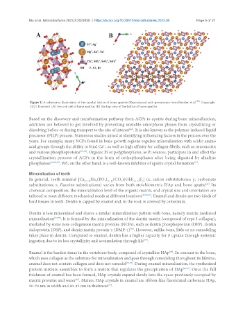
Mu et al. Microstructures 2023;3:2023030  https://dx.doi.org/10.20517/microstructures.2023.05  Page 5 of 21


                Figure 3. A schematic illustration of the crystal lattice of bone apatite (Reproduced with permission from Ressler  et al. [54] . Copyright
                2021, Elsevier). (A) the unit cell of bone apatite; (B) the top view of the lattice of bone apatite.

               Based on the discovery and transformation pathway from ACPs to apatite during bone mineralization,
               additives are believed to get involved by preventing unstable amorphous phases from crystallizing or
                                                                [42]
               dissolving before or during transport to the site of interest . It is also known as the polymer-induced liquid
               precursor (PILP) process. Numerous studies aimed at identifying influencing factors in the process over the
               years. For example, many NCPs found in bone growth regions regulate mineralization with acidic amino
               acid groups through the ability to bind Ca , as well as high affinity for collagen fibrils, such as osteonectin
                                                   2+
               and various phosphoproteins [66-68] . Organic Pi or polyphosphates, as Pi sources, participate in and affect the
               crystallization process of ACPs in the form of orthophosphates after being digested by alkaline
               phosphatase [32,69,70] . PPi, on the other hand, is a well-known inhibitor of apatite crystal formation .
                                                                                               [71]
               Mineralization of teeth
               In general, teeth mineral [Ca (10−x) Na (PO )  (CO )(OH) (2−z) z
                                                                     F ] (x, cation substitutions; y, carbonate
                                                    4 (6−y)
                                                            3
                                                x
                                                                                                     [40]
               substitutions; z, fluorine substitutions) varies from both stoichiometric HAp and bone apatite . Its
               chemical composition, the mineralization level of the organic matrix, and crystal size and orientation are
               tailored to meet different mechanical needs at different locations [37,72,73] . Enamel and dentin are two kinds of
               hard tissues in teeth. Dentin is capped by enamel and, in the root, is covered by cementum.
               Dentin is less mineralized and shares a similar mineralization pattern with bone, namely matrix-mediated
               mineralization [74,75] . It is formed by the mineralization of the dentin matrix (composed of type I collagen),
               mediated by some non-collagenous matrix proteins (NCPs), such as dentin phosphoprotein (DPP), dentin
               sialoprotein (DSP), and dentin matrix protein-1 (DMP-1) . However, unlike bone, little or no remodeling
                                                                [76]
               takes place in dentin. Compared to enamel, dentin has a higher capacity for F uptake through systemic
                                                                       [77]
               ingestion due to its less crystallinity and accumulation through life .

               Enamel is the hardest tissue in the vertebrate body, composed of crystalline HAp . In contrast to the bone,
                                                                                   [78]
               which uses collagen as the substrate for mineralization and goes through remodeling throughout its lifetime,
               enamel does not contain collagen and does not remodel [76,79] . During enamel mineralization, the synthesized
               protein mixture assembles to form a matrix that regulates the precipitation of HAp [80,81] . Once the full
               thickness of enamel has been formed, HAp crystals expand slowly into the space previously occupied by
               matrix proteins and water . Mature HAp crystals in enamel are ribbon-like fluoridated carbonate HAp,
                                      [82]
               50-70 nm in width and 20-25 nm in thickness .
                                                     [83]