
Page 290 - Read Online
P. 290
Zhou et al. Microstructures 2023;3:2023043 https://dx.doi.org/10.20517/microstructures.2023.38 Page 7 of 23
[39]
and corrosion . After treatment, these specimens can proceed with cryo-transfer [33,34,40] . However, this pre-
sharpening method is most effective when the materials being studied have uniform distribution and high
density of microstructural features of interest, such as nanosized precipitates or grain boundaries (GBs) in
fine-grain materials [33,34,40] . For specimens that require extracting a specific region of interest (ROI) from a
bulk specimen, it is essential to use a FIB fabrication method that is compatible with low temperatures,
often referred to as cryo-FIB [41-46] .
Cryo-FIB necessitates cooling not just the specimen stage but also the lift-out micromanipulator . Both
[39]
[36]
can be achieved by connecting a flexible copper cold band finger to the coolant . The process of FIB lift-
out involves trenching and extracting the ROI in a bar [Figure 4A1]. Subsequently, the bar is attached to the
micromanipulator, which requires using a gas injection system (GIS) and targeted electron beam deposition
to create the attachment or weld. Typically, this weld is made of platinum or carbon. After attachment to
the micromanipulator, the bar is removed from the trench and placed onto the pre-sharpened microposts
for further tip shaping.
However, working with both the specimen and the micromanipulator at cryogenic temperatures presents a
challenge. The GIS deposition cannot function as usual since the injected gas will non-selectively condense
onto the surfaces of both the specimen and the micromanipulator, hindering the site-specific attachment .
[22]
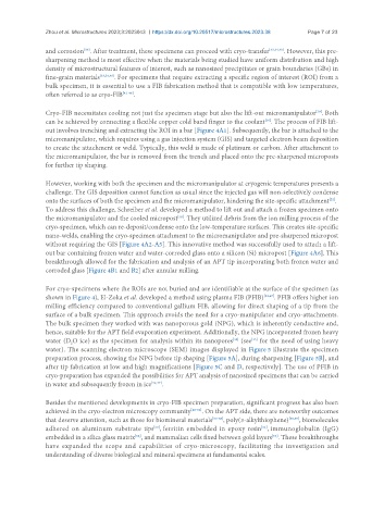
To address this challenge, Schreiber et al. developed a method to lift out and attach a frozen specimen onto
the micromanipulator and the cooled micropost . They utilized debris from the ion milling process of the
[39]
cryo-specimen, which can re-deposit/condense onto the low-temperature surfaces. This creates site-specific
nano-welds, enabling the cryo-specimen attachment to the micromanipulator and pre-sharpened micropost
without requiring the GIS [Figure 4A2-A5]. This innovative method was successfully used to attach a lift-
out bar containing frozen water and water-corroded glass onto a silicon (Si) micropost [Figure 4A6]. This
breakthrough allowed for the fabrication and analysis of an APT tip incorporating both frozen water and
corroded glass [Figure 4B1 and B2] after annular milling.
For cryo-specimens where the ROIs are not buried and are identifiable at the surface of the specimen (as
shown in Figure 4), El-Zoka et al. developed a method using plasma FIB (PFIB) [32,47] . PFIB offers higher ion
milling efficiency compared to conventional gallium FIB, allowing for direct shaping of a tip from the
surface of a bulk specimen. This approach avoids the need for a cryo-manipulator and cryo-attachments.
The bulk specimen they worked with was nanoporous gold (NPG), which is inherently conductive and,
hence, suitable for the APT field evaporation experiment. Additionally, the NPG incorporated frozen heavy
water (D O ice) as the specimen for analysis within its nanopores (see for the need of using heavy
[15]
[32]
2
water). The scanning electron microscope (SEM) images displayed in Figure 5 illustrate the specimen
preparation process, showing the NPG before tip shaping [Figure 5A], during sharpening [Figure 5B], and
after tip fabrication at low and high magnifications [Figure 5C and D, respectively]. The use of PFIB in
cryo-preparation has expanded the possibilities for APT analysis of nanosized specimens that can be carried
in water and subsequently frozen in ice [32,47] .
Besides the mentioned developments in cryo-FIB specimen preparation, significant progress has also been
achieved in the cryo-electron microscopy community [48-52] . On the APT side, there are noteworthy outcomes
that deserve attention, such as those for biomineral materials [53-58] , poly(3-alkylthiophene) [58,59] , biomolecules
[61]
adhered on aluminum substrate tips , ferritin embedded in epoxy resin , immunoglobulin (IgG)
[60]
embedded in a silica glass matrix , and mammalian cells fixed between gold layers . These breakthroughs
[63]
[62]
have expanded the scope and capabilities of cryo-microscopy, facilitating the investigation and
understanding of diverse biological and mineral specimens at fundamental scales.