Page 425 - Read Online
P. 425
Linger et al. Vessel Plus 2020;4:36  I  http://dx.doi.org/10.20517/2574-1209.2020.51                                                 Page 3 of 9


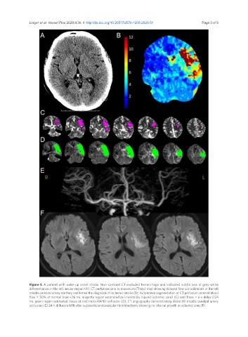
               Figure 1. A patient with wake-up onset stroke. Non-contrast CT excluded hemorrhage and indicated subtle loss of grey-white
               differentiation in the left insular region (A); CT perfusion time to maximum (Tmax) map showing delayed flow via collaterals in the left
               middle cerebral artery territory confirmed the diagnosis of ischemic stroke (B); Automated segmentation of CT perfusion cerebral blood
               flow < 30% of normal brain (36 mL magenta region estimated as irreversibly injured ischemic core) (C) and Tmax > 6-s delay (124
               mL green region estimated tissue at risk) from RAPID software (D); CT angiography demonstrating distal M1 middle cerebral artery
               occlusion (E) 24-h diffusion MRI after successful endovascular thrombectomy showing no interval growth in ischemic core (F)

