
Page 398 - Read Online
P. 398
Bader et al. Vessel Plus 2020;4:34 I http://dx.doi.org/10.20517/2574-1209.2020.36 Page 3 of 14
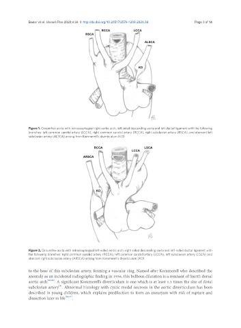
Figure 1. Circumflex aorta with retroesophageal right aortic arch, left sided descending aorta and left ductal ligament with the following
branches: left common carotid artery (LCCA), right common carotid artery (RCCA), right subclavian artery (RSCA), and aberrant left
subclavian artery (ALSCA) arising from Kommerell’s diverticulum (KD)
Figure 2. Circumflex aorta with retroesophageal left-sided aortic arch, right sided descending aorta and left-sided ductal ligament with
the following branches: right common carotid artery (RCCA), left common carotid artery (LCCA), left subclavian artery (LSCA) and
aberrant right subclavian artery (ARSCA) arising from Kommerell’s diverticulum (KD)
to the base of this subclavian artery, forming a vascular ring. Named after Kommerell who described the
anomaly as an incidental radiographic finding in 1936, this bulbous dilatation is a remnant of fourth dorsal
aortic arch [24-26] . A significant Kommerell’s diverticulum is one which is at least 1.5 times the size of distal
[4]
subclavian artery . Abnormal histology with cystic medal necrosis in the aortic diverticulum has been
described in young children, which explains predilection to form an aneurysm with risk of rupture and
dissection later in life [24,27] .