
Page 291 - Read Online
P. 291
Thirugnanam et al. Vessel Plus 2020;4:26 I http://dx.doi.org/10.20517/2574-1209.2020.18 Page 5 of 16
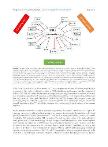
Figure 2. The role of SNRK in maintaining tissue homeostasis. Phosphorylation or signaling of SNRK to maintain homeostasis in three
systems - cardiac system, adipose system, and inflammatory system is shown. The arrow indicates whether the phosphorylation
is activating (black) or inhibiting (red) for the function of the target protein. The proteins under each system have been implicated
to communicate with SNRK in that system. SNRK: sucrose nonfermenting 1-related kinase; Trib3: tribbles homologue 3; PPARα:
peroxisome proliferator-activated receptor α; UCP3: uncoupling protein 3; ACC: Acetyl-coA carboxylase; AMPK: adenosine
monophosphate-activated protein kinase; ROCK: Rho-associated kinase; ERK: extracellular-signal-regulated kinase; IL-10: interleukin
10; NF-κB: nuclear factor kappa-light-chain-enhancer of activated B cells; IL-6: interleukin 6; TNF-α: tumor necrosis factor α; PRDM16:
PR domain containing 16; UCP1: uncoupling protein 1; PGC1α: peroxisome proliferator-activated receptor-γ co-activator 1α; PPP25RD:
protein phosphatase 2 regulatory subunit B’Delta; mTOR: mammalian target of rapamycin; AKT: protein kinase B
of ACC1 at S79 and ACC2 at S212 residue. ACC enzymes generates malonyl CoA from acetyl CoA (a
byproduct of FAO pathway). Phosphorylation of ACC is inhibitory and thus prevents the generation of
malonyl CoA. This relieves the inhibition on a transporter (carnitine palmitoyltransferase) that allows acyl
CoA to enter mitochondria for b-oxidation and tricarboxylic acid (TCA) cycle to generate acetyl CoA and
ATP respectively. In P0 Snrk global KO hearts, pAMPK and pACC levels were down compared to wild type
hearts suggesting malonyl CoA accumulation and thereby inhibition of carnitine palmitoyltransferase and
[15]
loss of b-oxidation or FAO . Thus, SNRK promotes FAO via the pAMPK-pACC pathway in the neonate
P0 hearts.
As the transition from the neonate to postnatal stage ensues, the heart of a newborn will adapt to the
changing environment which is characterized by an increase in contractile demand due to the rapid
[43]
growth and increase in activity of the newborn . This results in an increase in energy demand that can be
provided by FAO and mitochondrial ATP production. The high fatty acid content of the maternal milk in
many species is an effective way to supply the high energy demand of the newborn heart. As the newborn
heart matures, the utilization of FAO to meet the overall energy production needs increases, and thus
becomes the dominant substrate in the adult heart. Under normal metabolic conditions, over 95% of ATP
generated in the heart is derived from oxidative phosphorylation. Only 5% of the remaining comes from
glycolysis and to a lesser extent from the TCA cycle . Accordingly, the high-energy phosphate pool in the
[44]
heart, ATP, is relatively small and can be exhausted within a few seconds. Therefore, cardiac work depends