
Page 289 - Read Online
P. 289
Thirugnanam et al. Vessel Plus 2020;4:26 I http://dx.doi.org/10.20517/2574-1209.2020.18 Page 3 of 16
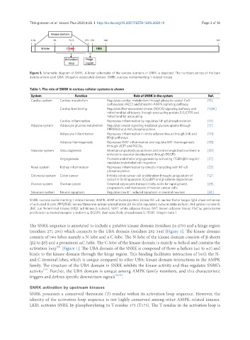
Figure 1. Schematic diagram of SNRK. A linear schematic of the various domains in SNRK is depicted. The numbers on top of the bars
denote amino acid. UBA: Ubiquitin-associated domain; SNRK: sucrose nonfermenting 1-related kinase
Table 1. The role of SNRK in various cellular systems is shown
System Function Role of SNRK in the system Ref.
Cardiac system Cardiac metabolism Regulates cardiac metabolism through phospho-acetyl-CoA [15]
carboxylase (ACC) and phospho-AMPK signaling pathway
Cardiac functioning Regulates Rho-associated kinase (ROCK) signaling pathway and [13,16]
mitochondrial efficiency through uncoupling protein 3 (UCP3) and
mitochondrial uncoupling
Cardiac inflammation Represses inflammation by regulates NF-κB phosphorylation [14]
Adipose system Adipocyte glucose metabolism Regulates insulin signaling mediated glucose uptake through [17]
PPP2R5D and Akt phosphorylation
Adipocyte inflammation Represses inflammation in white adipose tissue through JNK and [19]
IKKβ pathways
Adipose thermogenesis Represses WAT inflammation and regulate BAT thermogenesis [18]
through UCP1 and PGC1α
Vascular system Vasculogenesis Maintain angioblast populations and control angioblast numbers in [20]
embryonic vascular development through DUSP5
Angiogenesis Promote endothelial angiogenesis by activating ITGB1 (β1 integrin)- [21]
mediated endothelial cell migration
Renal system Kidney inflammation Represses inflammation by directly interacting with NF-κB [22]
phosphorylation
Colorectal system Colon cancer Inhibits colon cancer cell proliferation through upregulation of [12]
calcyclin-binding protein (CacyBP) and β-catenin degradation
Ovarian system Ovarian cancer Omental adipocytes transport fatty acids for rapid growth, [23]
progression, and metastasis of ovarian cancer cells
+
Neuronal system Neuron apoptosis Regulates low K - induced apoptosis in cerebral neurons [24]
SNRK: sucrose nonfermenting 1-related kinase; AMPK: AMP-activated protein kinase; NF-κB: nuclear factor kappa-light-chain-enhancer
of activated B cells; PPP2R5D: serine/threonine-protein phosphatase 2A 56 kDa regulatory subunit delta isoform; Akt: protein kinase-B;
JNK: Jun N-terminal kinase; IKKβ: IκB kinase β subunit; WAT: white adipose tissue; BAT: brown adipose tissue; PGC1α: peroxisome
proliferator-activated receptor γ isoform α; DUSP5: dual-specificity phosphatase 5; ITGB1: Integrin beta-1
The SNRK sequence is annotated to include a putative kinase domain (residues 24-270) and a hinge region
(residues 271-291) which connects to the UBA domain (residues 292-344) [Figure 1]. The kinase domain
consists of two lobes namely a N-lobe and a C-lobe. The N-lobe of the kinase domain consists of β-sheets
[β2 to β5] and a prominent αC helix. The C-lobe of the kinase domain is mainly α-helical and contains the
activation loop [Figure 1]. The UBA domain of the SNRK is composed of three α helices (α1 to α3) and
[10]
binds to the kinase domain through the hinge region. This binding facilitates interaction of both the N-
and C-terminal lobes, which is unique compared to other UBA: kinase domain interactions in the AMPK
family. The structure of the UBA domain in SNRK inhibits the kinase activity and thus regulates SNRK’s
activity . Further, the UBA domain is unique among AMPK family members, and this characteristic
[10]
triggers and defines specific downstream signals [26-28] .
SNRK activation by upstream kinases
SNRK possesses a conserved threonine (T) residue within its activation loop sequence. However, the
identity of the activation loop sequence is not highly conserved among other AMPK-related kinases.
LKB1 activates SNRK by phosphorylating its T-residue 173 (T173). The T residue in the activation loop is