
Page 73 - Read Online
P. 73
Zlatkina et al. Vessel Plus 2019;3:7 I http://dx.doi.org/10.20517/2574-1209.2019.03 Page 7 of 11
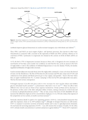
Figure 3. Intracellular mechanisms of insulin gene transcription damage by high concentrations of glucose and fatty acids[11]. ROS:
reactive oxygen radicals; PDX-1: pancreatic/duodenal homeobox-1; C/EBP β: CCAAT/enhancer binding protein β; JNK: C-jun N-terminal
kinase
[42]
synthesis) improves glucose homeostasis in insulin-resistant transgenic mice with obesity and diabetes .
Thus, PDX-1 and MafA are main targets of gluco- and lipotoxic processes, they operate in other ways.
Glucotoxicity is connected with a decrease in the expression of MafA and PDX-1 proteins, whereas in the
conditions of PDX-1 lipotoxicity it is expressed, but retained in the cytosol, the levels of MafA mRNA are
reduced.
At IR, the level of FFA in hepatocytes increases, because in these cells: (1) lipogenesis de novo increases; (2)
esterification of free fatty acids exceeds their oxidation; (3) esterified free fatty acids are stored in the form
of triglycerides or sent to the synthesis of cholesterol lipoproteins of very low density (VLDL); (4) insulin-
regulated mobilization of triglycerides decreases.
Insulin-resistant adipocytes intensely break down the triglycerides contained in them and release the formed
of them into the bloodstream. The flow of FFA from fat cells increases and FFA also comes out of VLDL and
chylomicrons from plasma and blood partially go to other organs, and partially - back to the liver where
they are again transformed into TG. A certain “pumping” of the liver by FFA and triglycerides takes place.
This has the most serious consequences.
Prolonged exposure of β-cells with fatty acids in vitro increases the main insulin release and inhibits
[43]
glucose-stimulated insulin secretion , a phenomenon observed in vivo in rats [44,45] . These two effects have
different time intervals and are likely to have separate mechanisms. Citrate-synthase activity decreases in
the presence of fatty acids in the islets cultivated within 24 hours, causing a decrease in citrate levels and
[46]
an increase in phosphofructokinase activity . This, in turn, reduces the levels of G6P, disinhibits GK and
increases the utilization of glucose at low concentrations of glucose. In vivo, FFA also reduces the response of
[47]
the sympathetic nervous system, so insulin secretion increases .
Particular attention should be paid to the separating protein-2 (UCP-2), a mitochondrial transporter that
[48]
splits the respiratory chain at the ATP synthesis stage , although its biological functions are still unclear.
UCP-2 is assumed to modulate secretion of hormone insulin and to enact in glucolipotoxicity formation.
On one hand, the increasing expression of UCP-2 - in β-cells weakens the secretion of insulin. On the other,
UCP-2 elevates insulin concentrations in animals and protects against diabetes [48,49] . Third, UCP-2 expression