
Page 364 - Read Online
P. 364
Page 6 of 10 Formica et al. Vessel Plus 2019;3:37 I http://dx.doi.org/10.20517/2574-1209.2019.19
A B
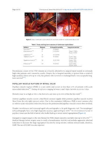
Figure 5. Apical ventricular septal defect (A); prosthetic patch for septal defect closure (B)
Table 2. Early and long-term outcome of ventricular septal defect
Authors Patients In-hospital mortality Long-term survival
Pojar et al. [44] 39 14 (36%) -
Okamoto et al. [45] 21 5 (23.8%) 3-year: 70.8%;
8-year: 57.9%
Liebelt et al. [42] 14 10 (71.4%) -
Takahashi et al. [46] 52 19 (36%) 5-year: 75%;
10-year: 31%
Papadopoulos et al. [47] 32 10 (31.2%) 5-year: 79%;
10-year: 51%
Prêtre et al. [48] 54 14 (26%) 5-year: 65%;
10-year: 40%
Percutaneous closure of the VSD remains an attractive alternative to surgical repair mainly in extremely
high-risks patients with reasonable results. Despite the in-hospital mortality is graved from a relatively
high incidence (from 20% up to 46%), the patients who survived to discharged had a very acceptable long-
term survival [43,49] .
PAPILLARY MUSCLE RUPTURE OF MITRAL VALVE
Papillary muscle rupture (PMR) is a rare entity and occurs in less than 0.5% of patients with acute
myocardial infarction . Timing of rupture is ranging between 2 and 7 days, but 80% occurs in 7 days.
[50]
Mortality may be as high as 50% in the first 24 h, and rises up to 90% within the first week .
[51]
Anterior papillary muscles receive a dual blood coronary supply, while posterior papillary muscle receives
blood from the only right coronary artery. Due to this anatomical difference, PMR is most common after
an inferior acute myocardial infarction, because the posteromedial papillary muscle is most often involved.
Doppler transthoracic and transesophageal echocardiography is the gold diagnostic tool. Transesophageal
[52]
echocardiography has a very high diagnostic accuracy approaching to 100% for the evidence of a tear in
papillary tissue and the flail of mitral valve leaflet leading to severe mitral regurgitation [Figure 6].
Emergent or urgent surgery is the only therapy for PMR, despite operative mortality rises up to 20%-39% [53-55] .
Medical therapy before surgery aims to reach a hemodynamic stability and includes aggressive afterload
reduction to decrease the huge regurgitant fraction by using nitrates, sodium nitroprusside, diuretics,
IABP. The use of ECMO was also reported.