
Page 291 - Read Online
P. 291
Page 4 of 18 Zimmermann et al. Vessel Plus 2019;3:31 I http://dx.doi.org/10.20517/2574-1209.2019.010
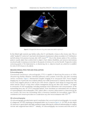
Figure 2. 2D Doppler Echo demonstrating atrial septal defects left-to-right shunt
by the dilated right ventricle may further reduce the LV end-diastolic volume in the chronic state. This so
called “masked LV restriction” may lead to development of pulmonary edema secondary to LV dysfunction
and left atrium (LA) pressure increase after ASD closure [22,25] . Due to the chronic nature of the condition,
patients usually adjust their activity level to adapt to their relative disabilities, and invasive interventions
are placed under increasing scrutiny due to the paucity of evidence for survival benefit. Prospective studies
evaluating quality of life improvements, or elucidating risk vs. objective benefit are called for to establish
the role of ASD closure in the elderly.
IMAGING MODALITIES FOR ASD EVALUATION
Echocardiography
Conventional transthoracic echocardiography (TTE) is capable of identifying the presence of ASDs,
characterizing chamber dilatation, estimated pulmonary artery pressure, shunt ratio, and other coexisting
cardiac conditions. Figure 2 demonstrates doppler imaging of an unrepaired ASD. Tissue doppler
imaging may be of particular use in elderly patients who suffer pronounced LV diastolic dysfunction. One
recent study suggests patients at risk for post ASD closure congestive heart failure by measuring early
[26]
mitral annular velocity to help direct volume management during and after ASD closure . In regard
to assessment of ASD morphology, including maximum defect dimensions and characterization of the
surrounding tissue rim, 2D TTE is somewhat limited. These limitations are surmounted with the adjunct
of transesophageal echocardiography (TEE) which offers a stepwise enhancement in characterizing the
size, location, and tissue rim surrounding ASDs to determine suitability for transcatheter repair. TEE is
considered a semi-invasive procedure so is undertaken only after initial evaluation with TTE [27,28] .
3D echocardiography
3D echocardiography provides better spatial visualization than conventional echocardiography. An example
of a diagnostic 3D TEE visualizing an unrepaired defect can be seen in Figure 3A. 3D TEE can also depict
3D structures in great detail with high-resolution images allowing for enhanced understanding of complex
[27]
valvular and congenital heart defects . Initially, 3D echocardiography was reconstructed from serial 2D