
Page 55 - Read Online
P. 55
Page 2 of 5 Wang et al. Vessel Plus 2018;2:7 I http://dx.doi.org/10.20517/2574-1209.2018.15
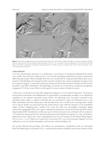
A B
C D
Figure 1. Initial selective angiography demonstrates flow through the proximal SMA (A) before redirection of contrast through the SMV (B)
and the venous system. After accessing the brachial artery, a 10 mm × 2.5 cm Viabahn stent was deployed (C) over the identified SMA
defect (via IVUS) successfully excluding the pathologic anatomy (D). SMA: superior mesenteric artery; SMV: superior mesenteric vein;
IVUS: intravascular ultrasound
CASE REPORT
A 44-year-old gentleman presented to us following a 4-year history of nonspecific abdominal discomfort
and a newly discovered peri-umbilical bruit. He recalled developing abdominal discomfort immediately
following laparoscopic Nissen fundoplication that was complicated by a large peritoneal bleed upon trocar
insertion. This bleeding event during the index operation resolved with copious application of surgical clips
by the general surgeon. A mesenteric duplex was performed which demonstrated turbulence in the superior
mesenteric vein (SMV) distribution. The pathologic connection was confirmed on a computed tomographic
angiogram (CTA), but it was difficult to distinguish the exact location of fistula formation.
At this point, we elected to proceed with a diagnostic angiogram via a transfemoral approach. The presence
and location of the fistula was established but no intervention was undertaken secondary to the significant
angulation of the superior mesenteric artery (SMA). The following week, the patient returned to the
operating theatre where a cutdown of the left arm was performed to expose the brachial artery to ease
SMA cannulation and stent deployment. The brachial artery was accessed with a micropuncture needle
and a long 6F sheath was positioned into the distal thoracic aorta with the assistance of an OmniFlush
angled catheter (AngioDynamics, Latham, NY). Once the SMA was successfully cannulated, digital
subtraction angiography (DSA), once again, confirmed an anomalous connection between the SMA and the
hypertrophied SMV; identification of the exact point of fistula formation was achieved with the assistance
of intravascular ultrasound (IVUS). The previously positioned 6F sheath was upsized to a long 12F sheath in
preparation of stent deployment. After exchanging the wire for a stiff Amplatz (Cook Medical, Bloomington,
IN), a 10 mm × 2.5 cm Viabahn self-expanding, covered stent (W.L. Gore and Associates, Flagstaff, AZ) was
deployed to cover the marked anomalous arteriovenous connection [Figure 1].
The patient tolerated the procedure well without any postoperative complications and was discharged
home the following day. His repeat CTA at 1-month postoperatively exhibited a patent stent, absence of any