
Page 26 - Read Online
P. 26
Page 2 of 5 Wang et al. Vessel Plus 2018;2:3. I http://dx.doi.org/10.20517/2574-1209.2017.38
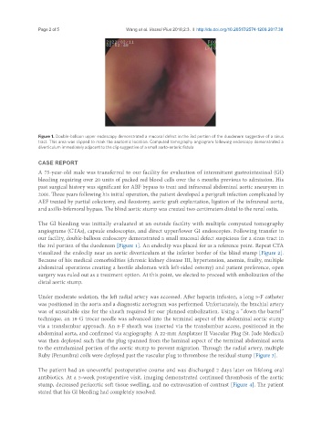
Figure 1. Double-balloon upper endoscopy demonstrated a mucosal defect in the 3rd portion of the duodenum suggestive of a sinus
tract. This area was clipped to mark the anatomic location. Computed tomography angiogram following endoscopy demonstrated a
diverticulum immediately adjacent to the clip suggestive of a small aorto-enteric fistula
CASE REPORT
A 75-year-old male was transferred to our facility for evaluation of intermittent gastrointestinal (GI)
bleeding requiring over 20 units of packed red blood cells over the 6 months previous to admission. His
past surgical history was significant for ABF bypass to treat and infrarenal abdominal aortic aneurysm in
2001. Three years following his initial operation, the patient developed a perigraft infection complicated by
AEF treated by partial colectomy, end ileostomy, aortic graft explantation, ligation of the infrarenal aorta,
and axillo-bifemoral bypass. The blind aortic stump was created two centimeters distal to the renal ostia.
The GI bleeding was initially evaluated at an outside facility with multiple computed tomography
angiograms (CTAs), capsule endoscopies, and direct upper/lower GI endoscopies. Following transfer to
our facility, double-balloon endoscopy demonstrated a small mucosal defect suspicious for a sinus tract in
the 3rd portion of the duodenum [Figure 1]. An endoclip was placed for as a reference point. Repeat CTA
visualized the endoclip near an aortic diverticulum at the inferior border of the blind stump [Figure 2].
Because of his medical comorbidities (chronic kidney disease III, hypertension, anemia, frailty, multiple
abdominal operations creating a hostile abdomen with left-sided ostomy) and patient preference, open
surgery was ruled out as a treatment option. At this point, we elected to proceed with embolization of the
distal aortic stump.
Under moderate sedation, the left radial artery was accessed. After heparin infusion, a long 5-F catheter
was positioned in the aorta and a diagnostic aortogram was performed. Unfortunately, the brachial artery
was of unsuitable size for the sheath required for our planned embolization. Using a “down the barrel”
technique, an 18-G trocar needle was advanced into the terminal aspect of the abdominal aortic stump
via a translumbar approach. An 8-F sheath was inserted via the translumbar access, positioned in the
abdominal aorta, and confirmed via angiography. A 22-mm Amplatzer II Vascular Plug (St. Jude Medical)
was then deployed such that the plug spanned from the luminal aspect of the terminal abdominal aorta
to the extraluminal portion of the aortic stump to prevent migration. Through the radial artery, multiple
Ruby (Penumbra) coils were deployed past the vascular plug to thrombose the residual stump [Figure 3].
The patient had an uneventful postoperative course and was discharged 2 days later on lifelong oral
antibiotics. At a 5-week postoperative visit, imaging demonstrated continued thrombosis of the aortic
stump, decreased periaortic soft tissue swelling, and no extravasation of contrast [Figure 4]. The patient
stated that his GI bleeding had completely resolved.