
Page 151 - Read Online
P. 151
Page 2 of 5 Kao et al. Vessel Plus 2018;2:18 I http://dx.doi.org/10.20517/2574-1209.2018.42
A B
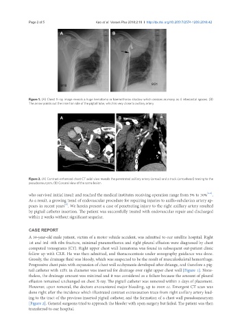
Figure 1. (A) Chest X-ray image reveals a huge hematoma or haemothorax shadow which crosses as many as 4 intercostal spaces. (B)
The arrow points out the insertion site of the pigtail tube, which is very close to axillary artery
A B
Figure 2. (A) Contrast-enhanced chest CT axial view reveals the penetrated axillary artery (arrow) and a track (arrowhead) tracing to the
pseudoaneurysm. (B) Coronal view of the same lesion
[4-6]
who survived initial insult and reached the medical institutes receiving operation range from 5% to 30% .
As a result, a growing trend of endovascular procedure for repairing injuries to axillo-subclavian artery ap-
pears in recent years . We herein present a case of penetrating injury to the right axillary artery resulted
[3]
by pigtail catheter insertion. The patient was successfully treated with endovascular repair and discharged
within 2 weeks without significant sequelae.
CASE REPORT
A 39-year-old male patient, victim of a motor vehicle accident, was admitted to our satellite hospital. Right
1st and 3rd -8th ribs fracture, minimal pneumothorax and right pleural effusion were diagnosed by chest
computed tomograms (CT). Right upper chest wall hematoma was found in subsequent out-patient clinic
follow up with CXR. He was then admitted, and thoracocentesis under sonography guidance was done.
Grossly, the drainage fluid was bloody, which was suspected to be the result of musculoskeletal hemorrhage.
Progressive chest pain with expansion of chest wall ecchymosis developed after driange, and therefore a pig-
tail catheter with 12Fr. in diameter was inserted for drainage over right upper chest wall [Figure 1]. None-
theless, the drainage amount was minimal and it was considered as a failure because the amount of pleural
effusion remained unchanged on chest X-ray. The pigtail catheter was removed within 3 days of placement.
However, upon removal, the doctors encountered major bleeding, up to 2000 cc. Emergent CT scan was
done right after the incidence which illustrated contrast extravasation trace from right axillary artery lead-
ing to the tract of the previous inserted pigtail catheter, and the formation of a chest wall pseudoaneurysm
[Figure 2]. General surgeons tried to approach the bleeder with open surgery but failed. The patient was then
transferred to our hospital.