
Page 48 - Read Online
P. 48
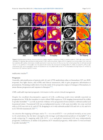
Pica et al. Vessel Plus 2022;6:10  https://dx.doi.org/10.20517/2574-1209.2021.81  Page 7 of 11


                Figure 3. Multiparameter tissue characterization by cardiac magnetic resonance (CMR) in amyloid patients. CMR with cines, native T1
                mapping, T2 mapping, late gadolinium enhancement (LGE), and extracellular volume (ECV) mapping in a normal subject (top row) and
                two patients with cardiac amyloidosis (middle and bottom rows) are shown. In the top row, the normal subject has no LGE and normal
                native T1, T2, and ECV. In the middle row, the patient with cardiac AL amyloidosis has high native T1, mildly increased T2, diffuse sub-
                endocardial LGE, and increased ECV values. In the bottom row, the patient with cardiac ATTR amyloidosis has high native T1, normal T2,
                transmural LGE, and very high ECV values.

               multicenter studies .
                               [44]

               Prognosis
               Currently, risk stratification of patients with AL and ATTR amyloidosis relies on biomarkers (NT-pro-BNP,
               troponin, free light chains, and eGFR) and clinical assessment, able to give prognostic information at
               presentation. Promising results derive from the study of the prognostic impact of changes of biomarkers, to
               assess disease progression and response to therapies [40,45] .


               CMR could add important prognostic information to the current clinical management.

               Despite the excellent discriminative capacity of LGE, conflicting results were initially reported on
               prognostication. With the transition to more robust PSIR sequences, LGE imaging demonstrated the ability
               to predict mortality [22,46] , as well as provide evidence of its progression from normal to subendocardial and
               transmural extent. Transmural LGE was an independent marker of all-cause mortality: two-year survival
               with no LGE was 92% in AL and ATTR, dropping to 81% with subendocardial LGE and 45% in AL and 65%
               in ATTR with transmural LGE [22,47] .

               As concerns the mapping technique, native T1 > 1044 ms and ECV > 0.45 predicted mortality at two years
               in AL amyloidosis, but the latter emerged as the stronger and independent predictor of mortality . In a
                                                                                                    [31]
               study comparing T1 mapping with LGE, ECV > 0.44 and global transmural LGE were independently
               prognostic; furthermore, in similar LGE patterns, ECV remained prognostic, while native T1 was not found
               to predict mortality [33,48] .