
Page 46 - Read Online
P. 46
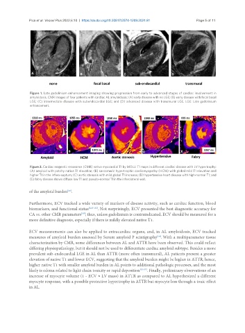
Pica et al. Vessel Plus 2022;6:10  https://dx.doi.org/10.20517/2574-1209.2021.81  Page 5 of 11


                Figure 1. Late gadolinium enhancement imaging showing progression from early to advanced stages of cardiac involvement in
                amyloidosis. CMR images of four patients with cardiac AL amyloidosis: (A) early disease with no LGE; (B) early disease with focal basal
                LGE; (C) intermediate disease with subendocardial LGE; and (D) advanced disease with transmural LGE. LGE: Late gadolinium
                enhancement.


                Figure 2. Cardiac magnetic resonance (CMR) native myocardial T1 by MOLLI T1 maps in different cardiac disease with LV hypertrophy:
                (A) amyloid with patchy native T1 elevation; (B) sarcomeric hypertrophic cardiomyopathy (HCM) with global mild T1 elevation and
                higher T1 in the infero-septum; (C) aortic stenosis with mild global T1 increase; (D) hypertensive heart disease with high-normal T1; and
                (E) fabry disease shows diffuse low T1 and pseudo-normal T1 in the inferolateral wall.

               of the amyloid burden .
                                  [20]

               Furthermore, ECV tracked a wide variety of markers of disease activity, such as cardiac function, blood
               biomarkers, and functional status [22,31,32] . Not surprisingly, ECV presented the best diagnostic accuracy for
               CA vs. other CMR parameters ; thus, unless gadolinium is contraindicated, ECV should be measured for a
                                         [33]
               more definitive diagnosis, especially if there is mildly elevated native T1.

               ECV measurements can also be applied to extracardiac organs, and, in AL amyloidosis, ECV tracked
               measures of amyloid burden assessed by Serum amyloid P scintigraphy . With a multiparameter tissue
                                                                             [34]
               characterization by CMR, some differences between AL and ATTR have been observed. This could reflect
               differing physiopathology, but it should not be used to differentiate cardiac amyloid subtype. Besides a more
               prevalent sub-endocardial LGE in AL than ATTR (more often transmural), AL patients present a greater
               elevation of native T1 and lower ECV, suggesting that the amyloid burden might be higher in ATTR; hence,
               higher native T1 with smaller amyloid burden in AL points to additional pathologic processes, and the most
               likely is edema related to light chain toxicity or rapid deposition [35-37] . Finally, preliminary observations of an
               increase of myocyte volume (1 - ECV × LV mass) in ATTR as compared to AL hypothesized a different
               myocyte response, with a possible protective hypertrophy in ATTR but myocyte loss through a toxic effect
               in AL.