


Page 26 - Read Online
P. 26
Smetanina et al. Vessel Plus 2021;5:19  https://dx.doi.org/10.20517/2574-1209.2021.17  Page 7 of 13


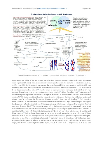
                      Figure 1. Schematic representation of the interplay of the genetic master regulators contributing to VVD development.

               investments and efforts of not one person, but collectives. However, without such data for veins (relative to
               other organs and tissues), further research on venous-specific action, for example, of certain medications,
               will be very difficult. Recently, it was shown that mitochondrial DNA copy number (mtDNA-CN) was
               inversely associated with incident and prevalent cardiovascular disease outcomes in 21,870 participants
               from three independent cohorts . Shortly after, in our laboratory, we found that mtDNA-CN was
                                            [36]
                                                                      [37]
                                                                                           [38]
               decreased in varicose vein vs. non-varicose vein tissue samples . Then, Castellani et al.  demonstrated
               across multiple independent cohorts that changes in mtDNA-CN influence nuclear DNA methylation at
               specific CpG loci and result in differential expression of specific genes that may impact human health and
               disease (namely, cardiovascular disease and all-cause mortality) via altered cell signaling . Understanding
                                                                                          [38]
               the mechanisms of mitochondrial and nuclear communication may shed light on the complex etiology of
               the disease, as well as the implications of therapeutic strategies to increase mitochondrial function. The fact
               that some drugs used in VVD treatment show their benefits in treating arterial diseases as well may serve as
               auxiliary evidence for the common molecular-genetic pathways in the pathogenesis of vascular diseases. A
               significant phlebotonic effect seems to be improbable without any impact on arterial smooth muscles and
                            [39]
               blood pressure . Indeed, horse chestnut extract contracts (through serotonin receptors, at least partly) both
                                                                        [40]
               veins and arteries, but it is more potent in inducing venocontraction . A pharmacological vasoactive agent,
               diosmin, is capable of inhibiting inflammatory pathways since it simultaneously influences pro-
               angiogenic/anti-angiogenic balance by an increase of anti-angiogenic factors and reducing the level of pro-
               angiogenic factors in blood plasma (TNF-alpha, VEGF-A and VEGF-C, angiostatin, IL-6, FGF2 and

