
Page 42 - Read Online
P. 42
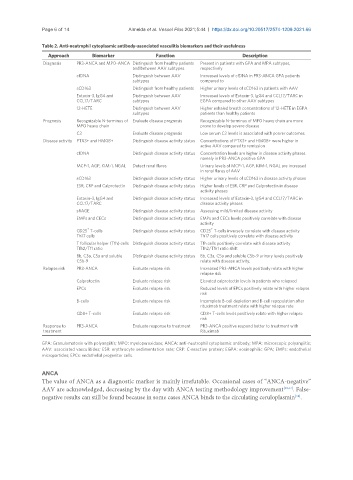
Page 6 of 14                 Almeida et al. Vessel Plus 2021;5:44  https://dx.doi.org/10.20517/2574-1209.2021.66

               Table 2. Anti-neutrophil cytoplasmic antibody-associated vasculitis biomarkers and their usefulness
                 Approach       Biomarker             Function                     Description
                Diagnosis  PR3-ANCA and MPO-ANCA Distinguish from healthy patients   Present in patients with GPA and MPA subtypes,
                                              and between AAV subtypes  respectively
                           cfDNA              Distinguish between AAV   Increased levels of cfDNA in PR3-ANCA GPA patients
                                              subtypes               compared to
                           sCD163             Distinguish from healthy patients  Higher urinary levels of sCD163 in patients with AAV
                           Eotaxin-3, IgG4 and   Distinguish between AAV   Increased levels of Eotaxin-3, IgG4 and CCL17/TARC in
                           CCL17/TARC         subtypes               EGPA compared to other AAV subtypes
                           12-HETE            Distinguish between AAV   Higher exhaled breath concentrations of 12-HETE in EGPA
                                              subtypes               patients than healthy patients
                Prognosis  Recognizable N-terminus of  Evaluate disease prognosis  Recognizable N-terminus of MPO heavy chain are more
                           MPO heavy chain                           prone to develop severe disease
                           C3                 Evaluate disease prognosis  Low serum C3 levels is associated with poorer outcomes
                Disease activity  PTX3+ and HMGB+  Distinguish disease activity status  Concentrations of PTX3+ and HMGB+ were higher in
                                                                     active AAV compared to remission
                           cfDNA              Distinguish disease activity status  Concentration levels are higher in disease activity phases
                                                                     namely in PR3-ANCA positive GPA
                           MCP-1, AGP, KIM-1, NGAL  Detect renal flares  Urinary levels of MCP-1, AGP, KIM-1, NGAL are increased
                                                                     in renal flares of AAV
                           sCD163             Distinguish disease activity status  Higher urinary levels of sCD163 in disease activity phases
                           ESR, CRP and Calprotectin  Distinguish disease activity status  Higher levels of ESR, CRP and Calprotectin in disease
                                                                     activity phases
                           Eotaxin-3, IgG4 and   Distinguish disease activity status  Increased levels of Eotaxin-3, IgG4 and CCL17/TARC in
                           CCL17/TARC                                disease activity phases
                           sRAGE              Distinguish disease activity status  Assessing mild/limited disease activity
                           EMPs and CECs      Distinguish disease activity status  EMPs and CECs levels positively correlate with disease
                                                                     activity
                              +                                         +
                           CD25  T-cells      Distinguish disease activity status  CD25  T-cells inversely correlate with disease activity
                           Th17 cells                                Th17 cells positively correlate with disease activity
                           T follicular helper (Tfh) cells  Distinguish disease activity status  Tfh cells positively correlate with disease activity
                           Tfh2/Tf1 ratio                            Tfh2/Tfh1 ratio shift
                           Bb, C3a, C5a and soluble   Distinguish disease activity status  Bb, C3a, C5a and soluble C5b-9 urinary levels positively
                           C5b-9                                     relate with disease activity.
                Relapse risk  PR3-ANCA        Evaluate relapse risk  Increased PR3-ANCA levels positively relate with higher
                                                                     relapse risk
                           Calprotectin       Evaluate relapse risk  Elevated calprotectin levels in patients who relapsed
                           EPCs               Evaluate relapse risk  Reduced levels of EPCs positively relate with higher relapse
                                                                     risk
                           B-cells            Evaluate relapse risk  Incomplete B-cell depletion and B-cell repopulation after
                                                                     rituximab treatment relate with higher relapse rate
                           CD8+ T-cells       Evaluate relapse risk  CD8+ T-cells levels positively relate with higher relapse
                                                                     risk
                Response to   PR3-ANCA        Evaluate response to treatment  PR3-ANCA positive respond better to treatment with
                treatment                                            Rituximab

               GPA: Granulomatosis with polyangiitis; MPO: myeloperoxidase; ANCA: anti-neutrophil cytoplasmic antibody; MPA: microscopic polyangiitis;
               AAV: associated vasculitides; ESR: erythrocyte sedimentation rate; CRP: C-reactive protein; EGPA: eosinophilic GPA; EMPs: endothelial
               microparticles; EPCs: endothelial progenitor cells.


               ANCA
               The value of ANCA as a diagnostic marker is mainly irrefutable. Occasional cases of “ANCA-negative”
               AAV are acknowledged, decreasing by the day with ANCA testing methodology improvement [36,37] . False-
               negative results can still be found because in some cases ANCA binds to the circulating ceruloplasmin .
                                                                                                    [38]