


Page 39 - Read Online
P. 39
Page 10 of 13          Echarte-Morales et al. Vessel Plus 2021;5:54  https://dx.doi.org/10.20517/2574-1209.2020.105


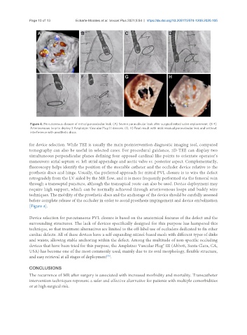
                Figure 4. Percutaneous closure of mitral paravalvular leak. (A) Severe paravalvular leak after surgical mitral valve replacement. (B-F)
                Arteriovenous loop to deploy 3 Amplatzer Vascular Plug III devices. (G, H) Final result with mild residual paravalvular leak and without
                interference with prosthetic discs.

               for device selection. While TEE is usually the main preintervention diagnostic imaging tool, computed
               tomography can also be useful in selected cases. For procedural guidance, 2D-TEE can display two
               simultaneous perpendicular planes defining four opposed cardinal-like points to orientate operator’s
               maneuvers: atrial septum vs. left atrial appendage and aortic valve vs. posterior aspect. Complementarily,
               fluoroscopy helps identify the position of the steerable catheter and the occluder device relative to the
               prothesis discs and hinge. Usually, the preferred approach for mitral PVL closure is to wire the defect
               retrogradely from the LV aided by the MR flow, and it is more frequently performed via the femoral vein
               through a transseptal puncture, although the transapical route can also be used. Device deployment may
               require high support, which can be normally achieved through arteriovenous loops and buddy wire
               techniques. The mobility of the prosthetic discs and the anchorage of the device should be carefully assessed
               before complete release of the occluder in order to avoid prosthesis impingement and device embolization
               [Figure 4].


               Device selection for percutaneous PVL closure is based on the anatomical features of the defect and the
               surrounding structures. The lack of devices specifically designed for this purpose has hampered this
               technique, so that treatment alternatives are limited to the off-label use of occluders dedicated to fix other
               cardiac defects. All of these devices have a self-expanding nitinol-based mesh with different types of disks
               and waists, allowing stable anchoring within the defect. Among the multitude of non-specific occluding
               devices that have been tried for this purpose, the Amplatzer Vascular Plug® III (Abbott, Santa Clara, CA,
               USA) has become one of the most commonly used, mainly due to its oval morphology, flexible structure,
               and easy retrieval at all stages of deployment .
                                                    [58]
               CONCLUSIONS
               The recurrence of MR after surgery is associated with increased morbidity and mortality. Transcatheter
               intervention techniques represent a safer and effective alternative for patients with multiple comorbidities
               or at high surgical risk.

