
Page 12 - Read Online
P. 12
Kovacs et al. Vessel Plus 2018;2:15 I http://dx.doi.org/10.20517/2574-1209.2018.06 Page 3 of 9
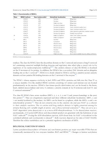
Table 1. Characterization of HDACs
Class HDAC isoform Size (amino acids) Subcellular localization Expression pattern
I HDAC1 482 Nucleus Ubiquitous
HDAC2 488 Nucleus Ubiquitous
HDAC3 428 Mainly nucleus Ubiquitous
HDAC8 377 Nucleus/cytoplasm Ubiquitous
IIa HDAC4 1084 Nucleus/cytoplasm Heart, skeletal muscle, brain
HDAC5 1122 Nucleus/cytoplasm Heart, skeletal muscle, brain
HDAC7 952 Nucleus/cytoplasm Heart, placenta, pancreas, skeletal muscle
HDAC9 1011 Nucleus/cytoplasm Skeletal muscle, brain
IIb HDAC6 1215 Mainly cytoplasm Heart, liver, kidney, pancreas
HDAC10 669 Mainly cytoplasm Liver, spleen, kidney
III SIRT1 747 Nucleus Ubiquitous
SIRT2 352 Cytoplasm Ubiquitous
SIRT3 399 Mitochondria Ubiquitous
SIRT4 314 Mitochondria Ubiquitous
SIRT5 310 Mitochondria Ubiquitous
SIRT6 355 Nucleus Ubiquitous
SIRT7 400 Nucleolus Ubiquitous
IV HDAC11 347 Mainly nucleus Brain, heart, skeletal muscle, kidney and testis
HDACs: histone deacetylases
residues. The class IIa HDACs have the deacetylase domain on the C-terminal and possess a long N-terminal
tail containing conserved multiple binding domains and regulatory sites which play a crucial role in the
regulation of the nucleocytoplasmic trafficking [29,33] . The catalytic domain of class IIb HDACs is localized
on the N-terminus of the protein. In addition, the HDAC6 has a secondary DAC domain and an ubiquitin
binding site on the C-terminal . HDAC10 is closely related to HDAC6 and has a putative second catalytic
[34]
[35]
domain and two putative Rb binding domains on the C-terminal of the enzyme .
The HDAC11 shares sequence similarity to both RPD3 and HDA1 proteins and falls into the Class IV as
a unique member. It is the smallest HDAC isoform consisting 347 amino acid residues and are located
predominantly in the nucleus. HDAC11 has tissue-specific distribution and can be found in the brain,
heart, skeletal muscle kidney and testis. It contains a catalytic domain at the N-terminus and short N- and
[36]
C-terminal extensions .
The class III HDACs have seven members (SIRT1, 2, 3, 4, 5, 6 and 7) and possess homology to the yeast
[37]
Sir2 protein . They are ubiquitously expressed and show different subcellular localization. SIRT1, 6 and
7 are mainly localized to the nucleus, the SIRT2 can be found in cytoplasm, while the SIRT3, 4 and 5 are
[38]
+
mitochondrial proteins . They do not comprise zinc in the catalytic site and uses NAD as a cofactor
in their catalytic reactions. The 275 amino acid long catalytic domain is highly conserved among the
[39]
sirtuins flanking with variable length of amino- and carboxy-terminal extensions . They can serve as a
+
NAD -dependent lysine deacetylase or as a mono-ADP-ribosyltransferase. In the deacetylation reaction,
nicotinamide, 2’-O-acetyl-ADP-ribose and deacetylated product are generated with the hydrolysis of one
+
[40]
+
NAD molecule . During the ADP-ribosylation reaction, ADP-ribose from the NAD is transferred to the
acetylated substrate and nicotinamide is released . Both reactions depend on the ratio of NAD /NADH.
[41]
+
Therefore the cellular metabolism can be a potential regulatory mechanism of SIRTs.
BIOLOGICAL FUNCTION OF HDACS
Lysine acetylation/deacetylation of histone and non-histone proteins are major reversible PTMs that are
[42]
dynamically maintained by two enzymes families, HAT and HDAC . Changes in histone acetylation