
Page 27 - Read Online
P. 27
Heng et al. Vessel Plus 2023;7:31 https://dx.doi.org/10.20517/2574-1209.2023.97 Page 5 of 14
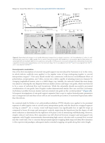
Figure 2. Biomechanical changes in vein grafts following transposition to arterial circulation. (A) Normalization of radius to wall
thickness ratio over time in rabbit jugular vein to carotid interposition grafts. (B) Similarities in sonographic spectral flow patterns in
unstented (left) and stented (right) porcine saphenous vein grafts. (C) Pressure to cross-sectional area ratio relationships in externally
supported and non-supported saphenous veins from domestic hogs. Part A was adapted from Ref. [16] ; part B was adapted from Ref. [15] ;
part C was adapted from Ref. [21] .
Hemodynamic modulation
One of the first descriptions of external vein graft support was introduced by Parsonnet et al. in the 1960s,
in which tubular scaffolds were applied to the jugular veins of dogs undergoing jugular to carotid
interposition surgery . This early sheath model was constructed with knitted monofilament fibers of
[20]
polyethylene, polypropylene, and Teflon, woven into a fabric capable of adjusting transverse diameter by
changing longitudinal tension, akin to a child’s finger trap. Initially, the rationale behind this stent design
was aimed at reducing the size disparity between smaller arteries and larger vein grafts, which was believed
to be a cause of turbulent flow and thrombosis. While the adjustable sheath successfully prevented
overdistention of vein grafts, later Doppler studies demonstrated similar flow rate and flow uniformity
(turbulence) profiles between stented and non-stented vein grafts at the carotid position [Figure 2B].
[15]
Subsequent investigations of vein graft support expanded their scope to explore hemodynamic parameters
such as tangential wall and shear stress with an aim towards the prevention of neointimal hyperplasia rather
than thrombosis.
In a seminal study by Kohler et al., polytetrafluoroethylene (PTFE) sheaths were applied to the proximal
segment of rabbit jugular vein to carotid artery interposition grafts, with the distal non-wrapped segment
serving as control . At 12 weeks, vessel wall hypertrophy was significantly less in tight-fit wraps as
[17]
compared to looser-fit wraps and unwrapped controls, supporting the hypothesis that the degree of wall
stress reduction as determined by the tightness of fit plays a role in attenuating wall thickening. However,
despite reduced wall stress, flow separation was still observed between wrapped and unwrapped vein
segments, with Doppler measurements demonstrating high central velocities and occasional flow reversal
near the vessel walls. Due to concerns that proximal segment wrapping likely impacted distal flow dynamics
in this experimental paradigm, subsequent studies extended wrapping vein grafts along their entire length.