
Page 73 - Read Online
P. 73
Page 14 of 29 Aguiar. Rare Dis Orphan Drugs J 2024;3:13 https://dx.doi.org/10.20517/rdodj.2023.56
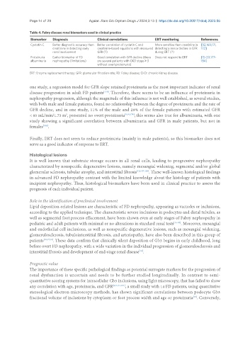
Table 4. Fabry disease renal biomarkers used in clinical practice
Biomarker Diagnosis Clinical correlations ERT monitoring References
Cystatin C Better diagnostic accuracy than Better correlation of cystatin C and More sensitive than creatinine in [92,169,171,
creatinine in detecting early creatinine-based equations with measured detecting a minor decline in GFR 172]
renal involvement GFR (?) during ERT (?)
Proteinuria Earlier biomarker of FD Good correlation with GFR decline (there Does not respond to ERT [15-20,177-
albuminuria nephropathy (limitations) are several patients with CKD stage ≥ 3 184]
without overt proteinuria)
ERT: Enzyme replacement therapy; GFR: glomerular filtration rate; FD: Fabry disease; CKD: chronic kidney disease.
one study, a regression model for GFR slope retained proteinuria as the most important indicator of renal
[179]
disease progression in adult FD patients . Therefore, there seems to be an influence of proteinuria in
nephropathy progression, although the magnitude of this influence is not well established, as several studies,
with both male and female patients, found no relationship between the degree of proteinuria and the rate of
GFR decline, and in one study, 11% of the male and 28% of the female patients with estimated GFR
< 60 mL/min/1.73 m , presented no overt proteinuria [9,10,178] ; this seems also true for albuminuria, with one
2
study showing a significant correlation between albuminuria and GFR in male patients, but not in
females .
[186]
Finally, ERT does not seem to reduce proteinuria (mainly in male patients), so this biomarker does not
serve as a good indicator of response to ERT.
Histological lesions
It is well known that substrate storage occurs in all renal cells, leading to progressive nephropathy
characterized by nonspecific degenerative lesions, namely mesangial widening, segmental and/or global
glomerular sclerosis, tubular atrophy, and interstitial fibrosis [15,187-190] . These well-known histological findings
in advanced FD nephropathy contrast with the limited knowledge about the histology of patients with
incipient nephropathy. Thus, histological biomarkers have been used in clinical practice to assess the
prognosis of each individual patient.
Role in the identification of preclinical involvement
Lipid deposition-related lesions are characteristic of FD nephropathy, appearing as vacuoles or inclusions,
according to the applied technique. The characteristic severe inclusions in podocytes and distal tubules, as
well as segmental foot process effacement, have been shown even at early stages of Fabry nephropathy in
pediatric and adult patients with minimal or no alterations in standard renal tests [15-20] . Moreover, mesangial
and endothelial cell inclusions, as well as nonspecific degenerative lesions, such as mesangial widening,
glomerulosclerosis, tubulointerstitial fibrosis, and arteriopathy, have also been described in this group of
patients [15,17,19] . These data confirm that clinically silent deposition of Gb3 begins in early childhood, long
before overt FD nephropathy, with a wide variation in the individual progression of glomerulosclerosis and
interstitial fibrosis and development of end-stage renal disease .
[12]
Prognostic value
The importance of these specific pathological findings as potential surrogate markers for the progression of
renal dysfunction is uncertain and needs to be further studied longitudinally. In contrast to semi-
quantitative scoring systems for intracellular Gb3 inclusions, using light microscopy, that has failed to show
any correlation with age, proteinuria, and GFR [9,15,17,191] , a small study with 14 FD patients, using quantitative
stereological electron microscopy methods, has shown significant correlations between podocyte Gb3
[16]
fractional volume of inclusions by cytoplasm or foot process width and age or proteinuria . Conversely,