
Page 789 - Read Online
P. 789
Pirakitikulr et al. Plast Aesthet Res 2020;7:67 I http://dx.doi.org/10.20517/2347-9264.2020.77 Page 3 of 10
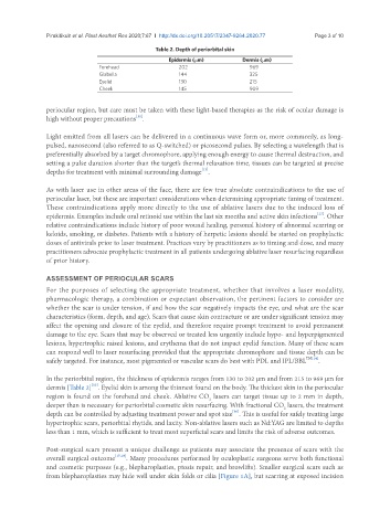
Table 2. Depth of periorbital skin
Epidermis (µm) Dermis (µm)
Forehead 202 969
Glabella 144 325
Eyelid 130 215
Cheek 145 909
periocular region, but care must be taken with these light-based therapies as the risk of ocular damage is
[11]
high without proper precautions .
Light emitted from all lasers can be delivered in a continuous wave form or, more commonly, as long-
pulsed, nanosecond (also referred to as Q-switched) or picosecond pulses. By selecting a wavelength that is
preferentially absorbed by a target chromophore, applying enough energy to cause thermal destruction, and
setting a pulse duration shorter than the target’s thermal relaxation time, tissues can be targeted at precise
depths for treatment with minimal surrounding damage .
[12]
As with laser use in other areas of the face, there are few true absolute contraindications to the use of
periocular laser, but these are important considerations when determining appropriate timing of treatment.
These contraindications apply more directly to the use of ablative lasers due to the induced loss of
[13]
epidermis. Examples include oral retinoid use within the last six months and active skin infections . Other
relative contraindications include history of poor wound healing, personal history of abnormal scarring or
keloids, smoking, or diabetes. Patients with a history of herpetic lesions should be started on prophylactic
doses of antivirals prior to laser treatment. Practices vary by practitioners as to timing and dose, and many
practitioners advocate prophylactic treatment in all patients undergoing ablative laser resurfacing regardless
of prior history.
ASSESSMENT OF PERIOCULAR SCARS
For the purposes of selecting the appropriate treatment, whether that involves a laser modality,
pharmacologic therapy, a combination or expectant observation, the pertinent factors to consider are
whether the scar is under tension, if and how the scar negatively impacts the eye, and what are the scar
characteristics (form, depth, and age). Scars that cause skin contracture or are under significant tension may
affect the opening and closure of the eyelid, and therefore require prompt treatment to avoid permanent
damage to the eye. Scars that may be observed or treated less urgently include hypo- and hyperpigmented
lesions, hypertrophic raised lesions, and erythema that do not impact eyelid function. Many of these scars
can respond well to laser resurfacing provided that the appropriate chromophore and tissue depth can be
safely targeted. For instance, most pigmented or vascular scars do best with PDL and IPL/BBL TM[14] .
In the periorbital region, the thickness of epidermis ranges from 130 to 202 µm and from 215 to 969 µm for
[15]
dermis [Table 2] . Eyelid skin is among the thinnest found on the body. The thickest skin in the periocular
region is found on the forehead and cheek. Ablative CO lasers can target tissue up to 2 mm in depth,
2
deeper than is necessary for periorbital cosmetic skin resurfacing. With fractional CO lasers, the treatment
2
[16]
depth can be controlled by adjusting treatment power and spot size . This is useful for safely treating large
hypertrophic scars, periorbital rhytids, and laxity. Non-ablative lasers such as Nd:YAG are limited to depths
less than 1 mm, which is sufficient to treat most superficial scars and limits the risk of adverse outcomes.
Post-surgical scars present a unique challenge as patients may associate the presence of scars with the
overall surgical outcome [17,18] . Many procedures performed by oculoplastic surgeons serve both functional
and cosmetic purposes (e.g., blepharoplasties, ptosis repair, and browlifts). Smaller surgical scars such as
from blepharoplasties may hide well under skin folds or cilia [Figure 1A], but scarring at exposed incision