
Page 601 - Read Online
P. 601
Page 2 of 9 Demzik et al. Plast Aesthet Res 2020;7:52 I http://dx.doi.org/10.20517/2347-9264.2020.93
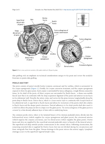
Figure 1. Penis cross sectional anatomy: (A) penile shaft and (B) penile glans
skin grafting with an emphasis on technical considerations unique to the penis and review the available
literature on penile skin grafting.
PENILE ANATOMY
The penis consists of paired erectile bodies (corpora cavernosa) and the urethra, which is surrounded by
the corpus spongiosum [Figure 1]. Distally, the corpus cavernosa terminate, and the corpus spongiosum
expands to form the glans penis. Each corpus is surrounded by tunica albuginea, a tough fibrous connective
tissue. In the shaft of the penis, all three corpora are surrounded by Buck’s fascia - a dense non-mobile
fascial layer that is in continuity with the deep suspensory ligament of the penis and anterior rectus fascia
in the abdominal wall. Dorsally, the artery, veins, and nerves for the glans traverse longitudinally along the
penis deep to Buck’s fascia. Dartos fascia, which is a loose areolar layer in continuity with Scarpa’s fascia of
the abdominal wall, is superficial to Buck’s fascia and allows for movement of the penile shaft skin relative
to Buck’s fascia and the deeper penile structures. Dermal adhesions in the distal penile shaft skin result in
a fold that allows the preputial skin to drape over the glans penis. The tunica albuginea of the glans penis is
covered by a thin densely adherent layer of skin with no underlying fascia.
The common penile artery, which is the terminal branch of the internal pudendal artery, divides into the
bulbourethral artery (which supplies the corpus spongiosum and glans penis), the cavernosal arteries
(which supplies the corpora cavernosa), and the dorsal artery (which supplies the glans penis). Dartos
fascia and skin are supplied by the superficial external pudendal artery (a branch of the femoral artery).
The superficial and deep arterial systems communicate at the coronal sulcus. Following circumcision, the
remaining preputial skin is supplied by Dartos fascia that was not divided during the circumcision and
from retrograde flow from the glans. Venous drainage in the penis mirrors the arterial supply. Lymphatic
drainage occurs via the superficial inguinal lymph nodes.