
Page 422 - Read Online
P. 422
Page 4 of 13 Singh et al. Plast Aesthet Res 2020;7:39 I http://dx.doi.org/10.20517/2347-9264.2019.76
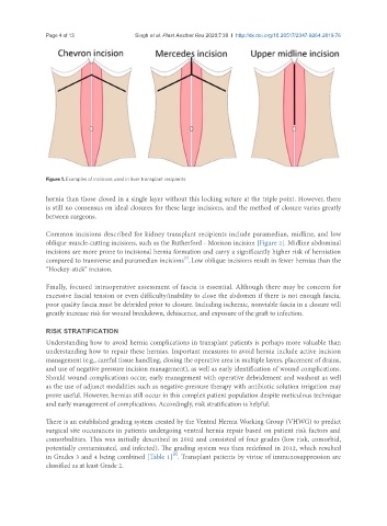
Figure 1. Examples of incisions used in liver transplant recipients
hernia than those closed in a single layer without this locking suture at the triple point. However, there
is still no consensus on ideal closures for these large incisions, and the method of closure varies greatly
between surgeons.
Common incisions described for kidney transplant recipients include paramedian, midline, and low
oblique muscle-cutting incisions, such as the Rutherford - Morison incision [Figure 2]. Midline abdominal
incisions are more prone to incisional hernia formation and carry a significantly higher risk of herniation
[2]
compared to transverse and paramedian incisions .Low oblique incisions result in fewer hernias than the
“Hockey-stick” incision.
Finally, focused intraoperative assessment of fascia is essential. Although there may be concern for
excessive fascial tension or even difficulty/inability to close the abdomen if there is not enough fascia,
poor quality fascia must be debrided prior to closure. Including ischemic, nonviable fascia in a closure will
greatly increase risk for wound breakdown, dehiscence, and exposure of the graft to infection.
RISK STRATIFICATION
Understanding how to avoid hernia complications in transplant patients is perhaps more valuable than
understanding how to repair these hernias. Important measures to avoid hernia include active incision
management (e.g., careful tissue handling, closing the operative area in multiple layers, placement of drains,
and use of negative pressure incision management), as well as early identification of wound complications.
Should wound complications occur, early management with operative debridement and washout as well
as the use of adjunct modalities such as negative-pressure therapy with antibiotic solution irrigation may
prove useful. However, hernias still occur in this complex patient population despite meticulous technique
and early management of complications. Accordingly, risk stratification is helpful.
There is an established grading system created by the Ventral Hernia Working Group (VHWG) to predict
surgical site occurances in patients undergoing ventral hernia repair based on patient risk factors and
comorbidities. This was initially described in 2002 and consisted of four grades (low risk, comorbid,
potentially contaminated, and infected). The grading system was then redefined in 2012, which resulted
[8]
in Grades 3 and 4 being combined [Table 1] . Transplant patients by virtue of immunosuppression are
classified as at least Grade 2.