
Page 414 - Read Online
P. 414
Ercin et al. Plast Aesthet Res 2020;7:38 I http://dx.doi.org/10.20517/2347-9264.2020.56 Page 3 of 7
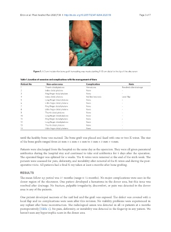
Figure 1. A 3 cm incision for bone graft harvesting was made starting 1-1.5 cm distal to the tip of the olecranon
Table 1. Location of nonunion and complications with the management of them
Patient No Non-union area Complication Note
1 Thumb distal phalanx Hematoma Resolved after drainage
2 Index distal phalanx None -
3 Ring Finger distal phalanx None -
4 Index distal phalanx Nail bed necrosis Local flap
5 Long Finger distal phalanx None -
6 Little finger distal phalanx None -
7 Ring Finger distal phalanx None -
8 Little finger distal phalanx None -
9 Thumb distal phalanx None -
10 Long Finger distal phalanx None -
11 Ring Finger distal phalanx None -
12 Long Finger distal phalanx None -
13 Thumb distal phalanx None -
14 Little finger distal phalanx None -
until the healthy bone was excised. The bone graft was placed and fixed with one or two K-wires. The size
of the bone grafts ranged from 20 mm × 6 mm × 6 mm to 3 mm × 3 mm × 4 mm.
Patients were discharged from the hospital on the same day as the operation. They were all given parenteral
antibiotics during the hospital stay and continued to take oral antibiotics for 5 days after the operation.
The operated finger was splinted for 6 weeks. The K-wires were removed at the end of the sixth week. The
patients were assessed for pain, deformity, and instability after removal of the K-wires and during the post-
operative visits. All patients had a final X-ray taken at least 6 months after bone grafting.
RESULTS
The mean follow-up period was 37 months (range 8-72 months). No major complications were seen in the
donor region of the olecranon. One patient developed a hematoma in the donor area, but this issue was
resolved after drainage. No fracture, palpable irregularity, discomfort, or pain was detected in the donor
area in any of the patients.
One patient developed necrosis of the nail bed and the graft was exposed. The defect was covered with a
local flap and no complications were seen after this revision. No viability problems were experienced in
any replant after bone reconstruction. The radiological union was detected in all 14 patients at 6 months
postoperatively [Table 1]. No pain, deformity, or instability was detected in the fingertip in any patient. We
haven’t seen any hypertrophic scars in the donor area.