
Page 254 - Read Online
P. 254
Page 6 of 11 Yamakawa et al. Plast Aesthet Res 2020;7:24 I http://dx.doi.org/10.20517/2347-9264.2020.20
A B
C D
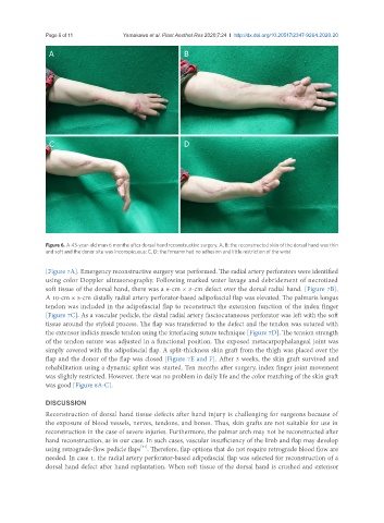
Figure 6. A 43-year-old man 6 months after dorsal hand reconstructive surgery. A, B: the reconstructed skin of the dorsal hand was thin
and soft and the donor site was inconspicuous; C, D: the forearm had no adhesion and little restriction of the wrist
[Figure 7A]. Emergency reconstructive surgery was performed. The radial artery perforators were identified
using color Doppler ultrasonography. Following marked water lavage and debridement of necrotized
soft tissue of the dorsal hand, there was a 6-cm × 3-cm defect over the dorsal radial hand. [Figure 7B].
A 10-cm × 5-cm distally radial artery perforator-based adipofascial flap was elevated. The palmaris longus
tendon was included in the adipofascial flap to reconstruct the extension function of the index finger
[Figure 7C]. As a vascular pedicle, the distal radial artery fasciocutaneous perforator was left with the soft
tissue around the styloid process. The flap was transferred to the defect and the tendon was sutured with
the extensor indicis muscle tendon using the interlacing suture technique [Figure 7D]. The tension strength
of the tendon suture was adjusted in a functional position. The exposed metacarpophalangeal joint was
simply covered with the adipofascial flap. A split-thickness skin graft from the thigh was placed over the
flap and the donor of the flap was closed [Figure 7E and F]. After 3 weeks, the skin graft survived and
rehabilitation using a dynamic splint was started. Ten months after surgery, index finger joint movement
was slightly restricted. However, there was no problem in daily life and the color matching of the skin graft
was good [Figure 8A-C].
DISCUSSION
Reconstruction of dorsal hand tissue defects after hand injury is challenging for surgeons because of
the exposure of blood vessels, nerves, tendons, and bones. Thus, skin grafts are not suitable for use in
reconstruction in the case of severe injuries. Furthermore, the palmar arch may not be reconstructed after
hand reconstruction, as in our case. In such cases, vascular insufficiency of the limb and flap may develop
[11]
using retrograde-flow pedicle flaps . Therefore, flap options that do not require retrograde blood flow are
needed. In case 1, the radial artery perforator-based adipofascial flap was selected for reconstruction of a
dorsal hand defect after hand replantation. When soft tissue of the dorsal hand is crushed and extensor