
Page 252 - Read Online
P. 252
Page 4 of 11 Yamakawa et al. Plast Aesthet Res 2020;7:24 I http://dx.doi.org/10.20517/2347-9264.2020.20
A B C
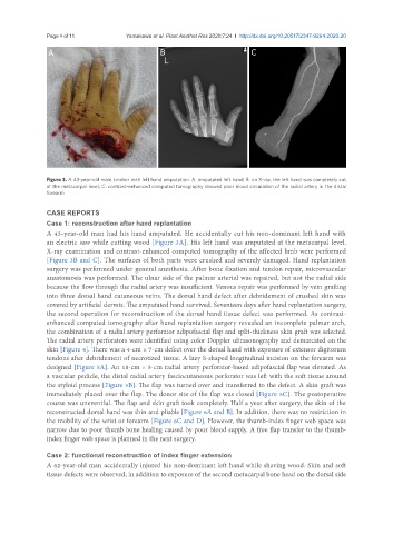
Figure 3. A 43-year-old male smoker with left hand amputation. A: amputated left hand; B: on X-ray, the left hand was completely cut
at the metacarpal level; C: contrast-enhanced computed tomography showed poor blood circulation of the radial artery in the distal
forearm
CASE REPORTS
Case 1: reconstruction after hand replantation
A 43-year-old man had his hand amputated. He accidentally cut his non-dominant left hand with
an electric saw while cutting wood [Figure 3A]. His left hand was amputated at the metacarpal level.
X-ray examination and contrast-enhanced computed tomography of the affected limb were performed
[Figure 3B and C]. The surfaces of both parts were crushed and severely damaged. Hand replantation
surgery was performed under general anesthesia. After bone fixation and tendon repair, microvascular
anastomosis was performed. The ulnar side of the palmar arterial was repaired, but not the radial side
because the flow through the radial artery was insufficient. Venous repair was performed by vein grafting
into three dorsal hand cutaneous veins. The dorsal hand defect after debridement of crushed skin was
covered by artificial dermis. The amputated hand survived. Seventeen days after hand replantation surgery,
the second operation for reconstruction of the dorsal hand tissue defect was performed. As contrast-
enhanced computed tomography after hand replantation surgery revealed an incomplete palmar arch,
the combination of a radial artery perforator adipofascial flap and split-thickness skin graft was selected.
The radial artery perforators were identified using color Doppler ultrasonography and demarcated on the
skin [Figure 4]. There was a 4-cm × 7-cm defect over the dorsal hand with exposure of extensor digitorum
tendons after debridement of necrotized tissue. A lazy S-shaped longitudinal incision on the forearm was
designed [Figure 5A]. An 18-cm × 5-cm radial artery perforator-based adipofascial flap was elevated. As
a vascular pedicle, the distal radial artery fasciocutaneous perforator was left with the soft tissue around
the styloid process [Figure 5B]. The flap was turned over and transferred to the defect. A skin graft was
immediately placed over the flap. The donor site of the flap was closed [Figure 5C]. The postoperative
course was uneventful. The flap and skin graft took completely. Half a year after surgery, the skin of the
reconstructed dorsal hand was thin and pliable [Figure 6A and B]. In addition, there was no restriction in
the mobility of the wrist or forearm [Figure 6C and D]. However, the thumb-index finger web space was
narrow due to poor thumb bone healing caused by poor blood supply. A free flap transfer to the thumb-
index finger web space is planned in the next surgery.
Case 2: functional reconstruction of index finger extension
A 62-year-old man accidentally injured his non-dominant left hand while shaving wood. Skin and soft
tissue defects were observed, in addition to exposure of the second metacarpal bone head on the dorsal side