
Page 304 - Read Online
P. 304
Nguyen et al. Plast Aesthet Res 2019;6:31 I http://dx.doi.org/10.20517/2347-9264.2019.42 Page 5 of 10
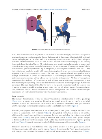
Figure 4. Quadriceps strengthening exercises post-reconstruction
at the time of initial resection. No patients had metastasis at the time of surgery. Two of the three patients
continue to survive despite metastatic disease that occurred at three years following initial treatment
in one, and eight years in the other. Both have pulmonary metastatic disease, and both have undergone
treatment for their metastasis, one in the form of Video-Assisted Thoracoscopic Surgery and the other by
Stereotactic Body Radiation Therapy. All patients underwent neoadjuvant radiation prior to surgery, and
none of the surviving patients received chemotherapy. The reconstructions following resection included a
free functional gracilis myocutaneous flap in one patient, a pedicled gracilis combined with an ALT flap
in 2 patients, and a pedicled gracilis with a deep inferior epigastric artery perforator/superficial inferior
epigastric artery (DIEP/SIEA) in one patient. The 3 surviving patients achieved MRC grade 4 muscle
strength and were able to achieve full knee extension 12-18 months post-operation. The three surviving
patients are able to ambulate without a brace and rise from the sitting position. All three of these patients
demonstrated clinical signs of re-innervation with palpable muscle contraction beginning at, or just
after 3 months post-operatively. Due to the clinical return of function, no electrophysiologic studies were
performed. The exact length of the obturator nerve to the gracilis was not recorded, but in every case
it was cut as short as possible to reduce re-innervation time yet still allow a tension free neurorrhaphy.
One patient died from his disease less than three months post-operation, and therefore it was too early to
demonstrate any contraction in the reinnervated functional reconstruction.
Case examples
The first case demonstrates a 50-year-old female with complete central resection of all four quadriceps
[Figure 5]. At 18 months post-operation, this patient has enough strength from her gracilis to reach full
extension, however she could not lock her knee into full extension for more than a short period of time
until more strength and endurance was achieved after several additional months [Figure 6].
Her anticipated progress is demonstrated in the following videos. MRC grade 1 strength with contraction
is noted at three months post-operation [Video 1]. MRC grade 2 contraction is noted at six months,
which provides enough knee stability to discontinue the knee brace and allow the patient to ascend and
descend stairs [Video 2]. MRC grade 3 strength is noted at nine months [Video 3]. MRC grade 4 strength
is demonstrated at one year from surgery, which enables the patient to return to more vigorous sporting
activities [Video 4]. Magnetic resonance imaging (MRI) demonstrates gracilis hypertrophy [Figure 7].
The second patient is a 22-year-old female with a large central resection of all four quadriceps muscles of
left leg, reconstructed with a free gracilis muscle. Figure 8 demonstrates her “neo” quadriceps function
with complete knee extension. She is able to complete a half marathon three years post-operation [Video 5].