
Page 302 - Read Online
P. 302
Nguyen et al. Plast Aesthet Res 2019;6:31 I http://dx.doi.org/10.20517/2347-9264.2019.42 Page 3 of 10
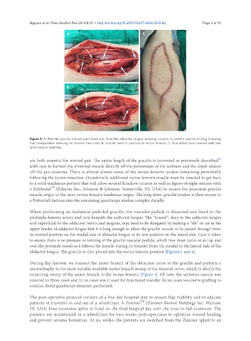
Figure 1. A: Pedicled gracilis harvest post-resection. Note the adductor longus releasing incision to prevent pedicle kinking following
flap transposition allowing for tension-free inset; B: Gracilis inset in position of rectus femoris; C: Skin defect was covered with free
anterolateral thigh flap
[5]
are both essential for normal gait. The entire length of the gracilis is harvested as previously described
with care to harvest the proximal muscle directly off the periosteum of the ischium and the distal tendon
off the pes anserine. There is almost always some of the rectus femoris tendon remaining proximally
following the tumor resection. Occasionally additional rectus femoris muscle must be resected to get back
to a solid tendinous portion that will allow several Krackow sutures as well as figure-of-eight sutures with
TM
0 Ethibond (Ethicon Inc., Johnson & Johnson, Somerville, NJ, USA) to secure the proximal gracilis
muscle origin to the stout rectus femoris tendinous origin. The long distal gracilis tendon is then woven in
a Pulvertaft fashion into the remaining quadriceps tendon complex distally.
When performing an ipsilateral pedicled gracilis, the vascular pedicle is dissected and freed to the
profunda femoris artery and vein beneath the adductor longus. The “tunnel”, deep to the adductor longus
and superficial to the adductor brevis and magnus, may need to be elongated by making a “slit” or cut in the
upper border of adductor longus that it is long enough to allow the gracilis muscle to be passed through from
its normal position on the medial side of abductor longus to its new position on the lateral side. Care is taken
to ensure there is no pressure or kinking of the gracilis vascular pedicle, which now must curve or arc up and
over the profunda vessels as it follows the muscle during its transfer from the medial to the lateral side of the
abductor longus. The gracilis is then placed into the rectus femoris position [Figures 1 and 2].
During flap harvest, we transect the motor branch of the obturator nerve to the gracilis and perform a
neurorrhaphy to the most suitable available motor branch stump of the femoral nerve, which is ideally the
remaining stump of the motor branch to the rectus femoris [Figure 3]. Of note, the sartorius muscle was
resected in three cases and in no cases was it used for functional transfer. In no cases was nerve grafting to
residual distal quadriceps elements performed.
The post-operative protocol consists of a five-day hospital stay to ensure flap viability and to educate
TM
patients in transfers in and out of a wheelchair. A Zimmer (Zimmer Biomet Holdings Inc. Warsaw,
IN, USA) knee extension splint is fitted on the final hospital day with the knee in full extension. The
patients are maintained in a wheelchair for two weeks post-operation to optimize wound healing
and prevent seroma formation. At six weeks, the patients are switched from the Zimmer splint to an