
Page 30 - Read Online
P. 30
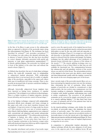
Figure 5: Sagittal view showing the prosthesis in-situ. Anterior to the   Figure 6: Postoperative anterior views of a young woman in (a) relaxed
           implant is the superior portion of pectoralis major. Posterior to the   position; (b) hands on hips; (c) arms fully abducted; and (d) forced
           implant lies pectoralis minor and the inferior portion of pectoralis major  contraction of pectoralis showing no dynamic muscle deformity

           in the line of its fibres to gain access to the submuscular   used to cover the superior pole of the implant has not been
           plane as opposed to division of the pectoralis major along   shown to cause any significant muscle contraction associated
           the infra-mammary fold [Figure 4]. The technique has been   deformities as may be the case with total sub muscular or
           described for primary [6-9]  and secondary procedures. [10-12]    dual plane techniques [Figure 6]. In comparison with partial
           The technique not only reduces the dynamic deformity due   sub muscular or dual plane implant positioning, where the
           to absence of muscle release but also has been described   muscle is released from the sternocostal margin, the biplane
           to correct dynamic deformity associated with partial sub   technique has the added  advantage of less incidences of
           muscular or dual plane augmentation  mammoplasty. [13,14]    dynamic breast deformity due to absence of the release of
           In Muscle splitting Biplane, the pectoralis lies behind and   the muscle. [4,13,14]  The muscle splitting technique does not
           in front of the implant at the same time and without the   require division of any of these fibres so that they are still
           muscle release [Figure 5].                          available for functional use.  The communication  between
                                                               the submuscular and sub glandular sections of the pocket
           As Tebbetts has described, the use of a dual plane technique   allows one unit feel of the breast. The sub glandular position
           reduces  the  trade-offs  commonly  seen  in  subglandular   of the implant in the lower pole also allows a more natural
           or subpectoral implant placement. With subglandular   and three-dimensional results with the implant covered by
           placements there is an increased risk of a visible or palpable   the muscle in the ever-changing upper part of the breast.
           edge of the prosthesis, especially in the upper pole where
           there may be insufficient soft tissue coverage. There is also   Intact sternal origin of the pectoralis muscle fibres acts as a
           possibly an increased risk of capsular contracture leading to   fence preventing the implant pockets join over the sternum,
           pain or breast deformity.                           thus, eliminating  the risk of synmastia.  When sternal
                                                               margins  of pectoralis are divided in  conventional or dual
           Although,  historically,  subpectoral breast  implants  have   plane pockets, the two pockets may communicate over the
           been reported as having lower incidences of capsular   sternum  resulting in synmastia. Subglandular positioning
           contracture,  the technique is not without its disadvantages.   of implant with medial quadrant undermining may result
                     [5]
           There is a higher incidence of implant migration, dynamic   in  similar complication.  The  correction  of sub  glandular
           breast deformity and less precise control of breast shape. [1]  synmastia can be corrected by simply converting the pocket
                                                               in to muscle splitting biplane.  To date there have been
                                                                                         [12]
           Use of the biplane technique compared with subglandular   no cases of synmastia and all of the patients have had an
           placement affords more adequate soft tissue  coverage in   aesthetically pleasing cleavage.
           the  upper pole with  a less stark transition  between  skin
           and implant. A long term review of a large study has shown   As the muscle-splitting technique  only divides the medial
           a 6-7 fold reduction in the over rate of revision surgeries,   two-thirds of pectoralis major, this  maintains  the lateral
           when Muscle Splitting Biplane augmentation was compared   portion of pectoralis major. The inferior retro-prosthetic
           with conventional sub glandular and partial submuscular   portion conjoins with the superior pre-prosthetic portion of
           augmentation mammoplasty.  The submuscular positioning   pectoralis major to locks the lateral part of the implant and
                                  [15]
           of implants in biplane also offers reduces incidence of   helps prevent superior and lateral displacement [Figure 4]. There
                            [5]
           capsular contracture.  In our series  there have been no   have not been any reported cases of implant displacement
           cases of capsular contracture so far, however, a larger series   or migration in our series.
           with well monitored long term follow up will be required for
           an actual rate of capsular contracture.             In comparison to submuscular implant placement, the
                                                               biplane technique affords the same adequacy of soft tissue
           The muscular attachment and portion of pectoralis major   cover in the superior pole, but in addition better fill and
           Plast Aesthet Res || Vol 3 || Issue 1 || Jan 15, 2016                                               19