
Page 29 - Read Online
P. 29
senior author and followed up prospectively. upward migration. The implant is inserted with the superior
portion in the subpectoral plane and the incision closed
Surgical technique: Midline is drawn from sternal notch occasionally with the placement of a drain.
to xiphisternum as a reference point and inframammary
incision is marked preoperatively with patient in standing RESULTS
position.
Follow up ranged from 9 months to 21 months. All of the
The procedure is performed in general anaesthetic with patients achieved precise and reliable implant placement
muscle relaxation with the patient in a supine position with no revisions or patient dissatisfaction. There have been
with their arms abducted. The marked mid-line is used for no cases of implant misplacement/migration; synmastia,
reference and future breast pocket is marked. Approximate dynamic breast deformity, capsular contracture or infections.
positions of the origins of pectoralis major are marked and A single case of unilateral haematoma occurred early in the
a line, extending between the junction of middle and lower series.
third of sternum and anterior axillary fold is drawn, roughly
level with the lower border of the areola. The line represents DISCUSSION
the level where the muscle splitting incision takes place. The
infra-mammary incisions are made approximately 5 cm in The use of a dual plane for breast augmentation has been
length and positioned laterally to conceal them in the infra- well documented in the past by Tebbetts. Dual plane is an
[1]
mammary fold [Figure 1]. extension of partial sub muscular technique where muscle
release is performed depending on the presence of the skin
Dissection first takes place in the sub-glandular plane using envelope. The bi-plane method, or muscle-splitting technique,
cutting diathermy and continues superiorly up to the level has been described by Khan in 2007. The submuscular
[4]
of the nipple-areola complex superiorly and between the positioning of the implant offers less capsular contracture
junction of middle and lower third of sternum medially rate. This method involves splitting the pectoralis major
[5]
going up and laterally to the anterior axillary fold [Figure 2].
The subpectoral pocket is accessed by separating the muscle
fibres close to their origin at the previously marked level
and the pocket is created by blunt dissection [Figure 3].
The medial two-thirds of pectoralis major are split in line
with the muscle fibres maintaining the lateral portion of the
muscle, which locks the implant and helps prevent lateral or
Figure 2: Arrows point to the level where the muscle-splitting incision
is made and lower unmarked area represents the extent of subglandular
pocket
Figure 1: Preoperative skin markings
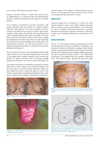
Figure 4: Anterior view showing position of the implant with the inferior
portion anterior to pectoralis major. The subpectoral plane is accessed by
splitting the muscle in the line of its fibres, lateral conjoined pectoralis
Figure 3: The muscle-splitting incision is made and access to the prevents lateral and superior displacements
subpectoral pocket is gained
18 Plast Aesthet Res || Vol 3 || Issue 1 || Jan 15, 2016