
Page 260 - Read Online
P. 260
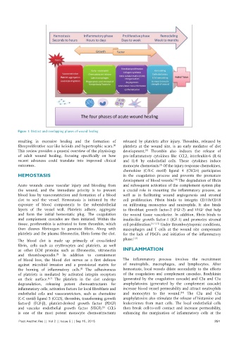
Figure 1: Distinct and overlapping phases of wound healing

          resulting in excessive healing and the formation of   released by platelets after injury. Thrombin, released by
                                                          [4]
          fibroproliferative scar‑like keloids and hypertrophic scars.    platelets at the wound site, is an early mediator of clot
          This review provides a general overview of the physiology   development.   Thrombin  also  induces  the  release  of
                                                                         [8]
          of adult wound healing,  focusing  specifically on how   pro‑inflammatory cytokines  like CCL2, interleukin‑6  (IL‑6)
          recent advances could translate  into improved clinical   and IL‑8 by endothelial cells. These cytokines induce
          outcomes.                                           monocyte chemotaxis.  Of the injury response chemokines,
                                                                                 [9]
                                                              chemokine  (C‑X‑C motif) ligand 4  (CXCL4) participates
          HEMOSTASIS                                          in  the  coagulation  process  and  prevents  the  premature
                                                              development of blood vessels.  The degradation of fibrin
                                                                                        [10]
          Acute wounds cause vascular injury and bleeding from   and subsequent activation of the complement system play
          the wound, and the immediate priority is to prevent   a  crucial role in mounting  the inflammatory  process, as
          blood loss  by  vasoconstriction  and formation  of a  blood   well  as in  facilitating wound  angiogenesis  and  stromal
          clot to seal the vessel.  Hemostasis  is  initiated  by  the   cell proliferation. Fibrin binds to integrin CD11b/CD18
          exposure  of blood components  to  the  subendothelial   on infiltrating monocytes and neutrophils. It also binds
          layers  of  the  vessel  wall. Platelets  adhere,  aggregate   to fibroblast growth factor‑2  (FGF‑2) and VEGF that help
          and form  the  initial  hemostatic  plug.  The  coagulation   the wound tissue vascularize. In addition, fibrin binds to
          and complement cascades are then initiated.  Within  the   insulin‑like growth factor‑1  (IGF‑1) and promotes stromal
          tissue, prothrombin is activated to form thrombin, which   cell proliferation. [5,11,12]  Under thrombocytopenic conditions,
          then  cleaves fibrinogen  to generate  fibrin.  Along with   macrophages and T cells at the wound site compensate
          platelets and the plasma fibronectin, fibrin forms the clot.  for the lack of PDGFs and initiation of the inflammatory
          The blood clot is made up primarily of cross‑linked   phase. [13]
          fibrin, cells such as erythrocytes and platelets, as well
          as other ECM proteins such as fibronectin, vitronectin   INFLAMMATION
          and  thrombospondin.   In  addition  to containment
                             [5]
          of blood loss, the blood clot serves as a first defense   The  inflammatory  process  involves  the  recruitment
          against microbial invasion and a provisional matrix for   of neutrophils, macrophages, and lymphocytes. After
          the homing of inflammatory cells.  The adhesiveness   hemostasis,  local vessels dilate secondarily to the effects
                                         [5]
          of platelets is mediated by activated integrin receptors   of the coagulation and  complement cascades.  Bradykinin
          on their surface. [6,7]  The platelets in the clot undergo   (generated by  the  coagulation cascade) and C3a and C5a
          degranulation, releasing potent chemoattractants for   anaphylatoxins (generated by the complement cascade)
          inflammatory cells, activation factors for local fibroblasts and   increase blood vessel permeability and attract neutrophils
          endothelial  cells  and  vasoconstrictors,  such  as  chemokine   and monocytes to the wound.  The C3a and C5a
                                                                                           [14]
          (C‑C motif) ligand 5 (CCL5), thrombin, transforming growth   anaphylatoxins also stimulate the release of histamine and
          factor‑b (TGF‑b), platelet‑derived growth  factor  (PDGF)   leukotrienes  from  mast  cells. The  local endothelial cells
          and vascular endothelial growth factor (VEGF).  CCL5   then break cell‑to‑cell contact and increase permeability,
                                                    [5]
          is one of the most potent monocyte chemoattractants   enhancing  the  margination  of inflammatory cells at the
          Plast Aesthet Res || Vol 2 || Issue 5 || Sep 15, 2015                                             251