
Page 145 - Read Online
P. 145
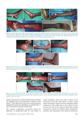
a                       b                        c          d


           e                                                  f                                       g
          Figure 1: (a) Crush injury to the right heel pad and ankle regions with multiple lacerations on the medial aspect of the foot; (b) crush injury to the
          right heel pad and ankle regions with multiple lacerations on the lateral aspect of the foot; (c) picture showing necrotic heel pad tissue; (d) extended
          reverse sural artery flap was elevated on day 2 following debridement; (e) extended reverse sural artery flap delay performed; (f) flap inset was
          completed on day 4 following debridement; (g) postoperative day 21 following surgery-posterior view


                         a                       b                       c


                                      d                       e
          Figure 2: (a) Ulceration on the lateral malleolus with exposure of the ankle joint; (b) picture following wound debridement and planning of a lateral
          supramalleolar flap; (c) lateral supramalleolar flap was elevated on day 2 following debridement; (d) lateral supramalleolar flap delay performed; (e) flap
          inset was completed on day 4 following debridement


                        a                     b                       c


                                       d                       e
          Figure 3: (a) Posttraumatic soft tissue defect exposing the lower third of the tibia; (b) distally-based posterior tibial artery perforator plus flap elevated;
          (c) distally-based  posterior tibial  artery perforator plus flap delay performed; (d) flap inset was completed on day 4 following debridement;  (e) late
          postoperative picture of flap

          based on the peroneal or posterior tibial artery perforators   venous congestion, which may result in failure at the
          are frequently used for reconstruction of defects in this   distal aspect of the flap, which may be covering a critical
          region. [1-3]  These flaps may be harvested as fasciocutaneous,   region of the defect. Causes for venous congestion include
          islanded fasciocutaneous, adipofascial, or propeller flaps, or   compression of the pedicle due to poor  elasticity of the
          may be harvested as delayed extended flaps.         skin over the  roof of the  tunnel  in  island flaps, valvular
          One  common complication encountered during the     incompetence, edema at the pedicle region, compartment
          utilization  of distally-based flaps for such defects is   syndrome, and compression of the pedicle by hematoma.

          Plast Aesthet Res || Vol 2 || Issue 3 || May 15, 2015                                             135