
Page 15 - Read Online
P. 15
BMP-2 and BMP-7 have also been reported to promote
bone consolidation during DO. Therefore, exogenous
[47]
administration of BMP may enhance DO both temporally
and spatially and enable rapid distraction, thereby
shortening the time to repair the bone defect. Although
various biomaterials have been used as injectable
delivery systems in DO models, little has been reported
on the use of nanobiomaterials as carrier materials
for the sustained release of growth factors in bone
regeneration.
The most widely explored osteogenic factors are the
members of the transforming BMP-2 family, which have
all been shown to augment the bone-forming capacity
of osteoblastic cell populations when delivered at the
[48]
appropriate times in the wound-healing cascade.
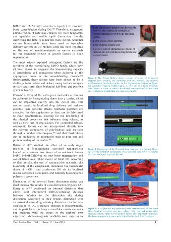
Unfortunately, these factors have been shown to be a Figure 2: The Phenix M-Bone device consists of a pair of permanent
magnets (one external, the controller, and one internal, the receiver,
challenge to formulate and deliver, owing to their complex which transmit mechanical power to the implanted device) upper figure:
tertiary structures, short biological half-lives, and possible the controller magnet; middle figure: a lever arm as a force amplifier;
systemic toxicity. lower figure: a screw to convert alternating movements of the lever arm
into continuous longitudinal one-way movements
Efficient delivery of the osteogenic molecules in vivo can
be achieved by incorporating them into a carrier, which
can be implanted directly into the defect site. This
method results in localized drug delivery and reduces
possible toxic systemic effects. Synthetic polymers are
attractive for this application as they can be fabricated
to exact specifications, allowing for the fine-tuning of
the physical properties that influence drug release, as
well as their rate of degradation. For controlled release,
osteogenic factors can be incorporated directly into
the polymer component of poly-hydroxy acid matrices
through a number of techniques, and their final release
[49]
can be modulated by parameters such as pore size and
protein loading of the matrix. [50]
Haidar et al. studied the effect of an early single
[50]
injection of biodegradable core-shell nanoparticles Figure 3: Photograph of the Phenix-M bone transport rod (above). Close-
loaded with various low doses of recombinant human up of bone transport mechanism, with threaded core used to transfix
BMP-7 (rhBMP-7/rhOP-1) on new bone regeneration and the bone transport segment (below)
consolidation in a rabbit model of tibial DO. According
to their results, the use of nanoparticles maintains the
bioactivity of the encapsulant, minimizes the therapeutic
doses of rhOP-1, and accelerates DO via its localized
release-controlled osteogenic, and naturally biocompatible
polymeric properties.
Elimination of the external frame distraction device can
itself improve the results of osteodistraction [Figures 2-5].
Konaş et al. developed an internal distractor that
[51]
allows local intermittent BMP-2-containing chitosan
hydrogel infusion to the distraction site during
distraction. According to their results, distraction with
an osteoinductive drug-releasing distractor can increase
ossification in DO. Moreover, chitosan is biocompatible,
and its particles act as bony extracellular matrix elements Figure 4: A 15-year-old boy presented with osteosarcoma of the distal
and integrate with the tissue. In the authors’ own tibia. Surgical resection specimen (above, left). Surgical defect after
resection (above, right). Bone transport device, after implantation (below).
experience, chitosan–alginate scaffolds were superior to The bone transport segment can be identified in the left of the figure
Plast Aesthet Res || Vol 1 || Issue 1 || Jun 2014 9