
Page 13 - Read Online
P. 13
Page 8 of 9 Shetty. Plast Aesthet Res 2022;9:47 https://dx.doi.org/10.20517/2347-9264.2022.41
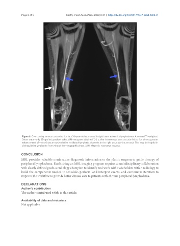
Figure 5. Overcoming venous contamination in a 70-year-old woman with right lower extremity lymphedema. A coronal T1-weighted
Dixon water-only 3D spoiled gradient echo MRI venogram obtained 120 s after intravenous contrast administration shows greater
enhancement of veins (blue arrows) relative to dilated lymphatic channels in the right ankle (white arrows). This may be helpful in
distinguishing lymphatics from veins at the venographic phase. MRI: Magnetic resonance imaging.
CONCLUSION
MRL provides valuable noninvasive diagnostic information to the plastic surgeon to guide therapy of
peripheral lymphedema. Establishing an MRL imaging program requires a multidisciplinary collaboration
with clearly defined goals, a radiology champion to identify and work with stakeholders within radiology to
build the components needed to schedule, perform, and interpret exams, and continuous iteration to
improve the workflow to provide better clinical care to patients with chronic peripheral lymphedema.
DECLARATIONS
Author’s contribution
The author contributed solely to this article.
Availability of data and materials
Not applicable.